<span>Stark Neurosciences Research Institute recently&nbsp;</span>formed three new research interest groups: ocular neurobiology,&nbsp;neurodevelopment and neuroimaging (in vivo). The more than 100 members of the institute&mdash;which include researchers from multiple departments at IU School of Medicine and IUPUI&mdash;can self-identify with one or more of the eight neuroscience-related research groups.

Stark Neurosciences adds to its research interests

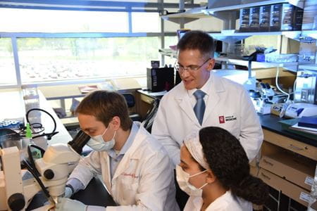
Tim Corson, PhD, works with medical students doing research in the lab.

Tim Corson, PhD, works with medical students doing research in the lab.

The human eye is the most visible part of the central nervous system—not only supplying a person with the sense of sight but functioning as an integral connection to the brain.

Researchers at Indiana University School of Medicine—combining the efforts and expertise of Stark Neurosciences Research Institute and the Eugene and Marilyn Glick Eye Institute—are studying how neurodegenerative diseases, such as Alzheimer’s and Parkinson’s diseases, age-related macular degeneration, and glaucoma, may manifest in the eye’s retina.

Several of these investigators are part of the newly formed ocular neurobiology research interest group at Stark Neurosciences Research Institute. The institute also established neurodevelopment and neuroimaging (in vivo) as new interest groups, which join addiction, neurodegenerative disorders, pain, psychiatric disorders and spinal cord and brain injury.

The more than 100 members of Stark Neurosciences Research Institute—which include researchers from multiple departments at IU School of Medicine and IUPUI—can self-identify their research interests within one or more of the eight groups.

Ocular Neurobiology

Some of the newest members of Stark Neurosciences Research Institute have come from the Department of Ophthalmology and Eugene and Marilyn Glick Eye Institute.

Over the past few years, the Department of Ophthalmology has more than doubled its basic and translational research division, and each of those scientists new to the department have joined or are in the process of joining Stark Neurosciences Research Institute as primary or affiliate members.

20977-Corson, TimothyTim Corson, PhD, associate professor of ophthalmology, has been a member of Stark Neurosciences Research Institute since 2018. His lab focuses on investigating neovascular eye diseases caused by abnormal blood vessel growth in the eye that can lead to retinal cell death.

“It’s a really great opportunity that we’ve been able to bring Glick Eye Institute researchers into Stark to build our nucleus,” Corson said, “and we look forward to future collaborations—as we grow and Stark grows, there’s only going to be more opportunities for people from the eye side of things to look at brain diseases.”

The major research focus of the ocular neurobiology group is the degeneration of different cell types in the eye’s retina. Approximately 90 percent of external information conveyed to the brain is visual and generated in the eye. And more than 15 million Americans suffer from ocular neurodegenerative diseases, such as age-related macular degeneration, glaucoma and diabetic retinopathy.

The retina, a thin layer of tissue located in the back of the eye, senses light and sends images to the brain through the optic nerve. It also serves as a window into the central nervous system to study disease progression of neurodegeneration, researchers say.

48163-Imanishi, YoshikazuYoshikazu Imanishi, PhD, associate professor of ophthalmology, has been studying sensory neurobiology since he was a graduate student. His lab develops novel small molecule therapies for inherited blinding disorders caused by degeneration of photoreceptor neurons.

Imanishi, a researcher at Glick Eye Institute since summer 2020, became a member of Stark Neurosciences Research Institute and joined the ocular neurobiology interest group later that year in order to collaborate with neuroscience investigators and train graduate students who are interested in ocular neurobiology, he said.

25526-Meyer, Jason

Jason Meyer, PhD, associate professor of medical and molecular genetics, investigates retinal ganglion cells. When these cells—which serve as the connection between the eye and the brain—degenerate through glaucoma, it leads to the loss of vision and eventual blindness.

Recent research has shown the retina—which can be seen in a standard ophthalmic exam—can be used as a powerful tool in the study of neurodegenerative diseases, Meyer said. Shannon Risacher, PhD, assistant professor of radiology and imaging services and part of the ocular neurobiology interest group, published a study last year on this phenomenon.

 “In the case of neurodegenerative diseases such as Alzheimer’s disease or Parkinson’s disease, it is thought that the eye provides a minimally invasive window to visualize the progression of neurodegeneration within the nervous system,” Meyer said, “leading to earlier diagnoses and the ability to develop personalized medicine approaches based upon ophthalmic examinations.”

Neuroimaging

The neuroimaging (in vivo) research interest group is part of an already strong imaging environment at IU School of Medicine. Many of its investigators are involved with the Indiana Institute of Biomedical Imaging Sciences (IIBIS) and the Center for Neuroimaging (CfN).

6553-Yoder, Karmen

Karmen Yoder, PhD, professor of radiology and imaging sciences, said neuroimaging at IU School of Medicine is a collaborative effort in and of itself, adding that the Center for Neuroimaging—located within Goodman Hall—being connected by a hallway to dozens of laboratories at Stark Neurosciences Research Institute takes this collaboration to the next level.

“Not only do we have opportunities to interact with outstanding basic research scientists who inform our work, we also have tremendous potential for truly translational work between the preclinical and human imaging cores,” Yoder said. “This is a really unique arrangement, and I am looking forward to seeing the groundbreaking neuroscience that is emerging here.”

One of the newest pieces of imaging equipment at IU School of Medicine is the Bruker BioSpec 9.4T PET-MRI scanner, located in the Roberts Translational Imaging Facility at Stark Neurosciences Research Institute—the second of its kind in the world. This 25,000-pound machine provides simultaneous MRI and PET imaging on small animal models, which allows for researchers to map animal model disease progression and therapeutic responses.

The PET-MRI scanner and other cutting-edge imaging technologies at IU School of Medicine are used to study a variety of neurological and psychiatric disorders, including normative aging and neurodegenerative diseases (Alzheimer’s disease), alcohol and substance use disorders, traumatic brain injury and adolescent and adult neuropsychiatric disorders.

Neurodevelopment

The neurodevelopment research interest group includes investigators who conduct preclinical or clinical research at IU School of Medicine.

Much of the preclinical research among faculty in the group is derived from basic science and developmental neurobiology research programs at the school. Investigators have a variety of neurodevelopment research interests, including molecular mechanisms guiding cell fate differentiation, axon guidance and synaptogenesis, as well as using pluripotent stem cells to produce sensory neurons, inner ear hair cells and retinal ganglion cells.

Brian Pierchala, PhD, Sherry Sonneborn Professor of ALS Research, is one of the many investigators who conduct preclinical neurodevelopment research. His lab investigates the development and regenerative capacity of peripheral axons, such as motor axons, with a focus on motor neuron degeneration and death due to amyotrophic lateral sclerosis (ALS).

Clinical neurodevelopment research focuses on understanding how differences in structure and function of the developing human brain affect cognition, behavior and psychosocial outcomes. Two members of the neurodevelopment group interest primarily on this type of clinical research.

Brenna McDonald, PsyD, professor of radiology and imaging sciences, has studied mild traumatic brain injury/concussion in children and adolescents, cognitive and brain effects of leukemia chemotherapy and brain structure and function in kids with temporal lobe seizures. Leslie Hulvershorn, MD, associate professor of psychiatry, investigates child and adolescent mental health and addiction. These studies have used neuroimaging to better understand brain-behavior relationships and to examine the neurobiological basis of emotion regulation and addiction risk.

Default Author Avatar IUSM Logo
Author

Ben Middelkamp

Ben Middelkamp is a communications manager for the Department of Neurology, Department of Neurological Surgery and Stark Neurosciences Research Institute at Indiana University School of Medicine. Before joining the Office of Strategic Communications in December 2019, Ben spent nearly six years as a newspaper reporter in two Indiana cities. He earned a bachelor’s degree in Convergent Journalism from Indiana Wesleyan University in 2014. Ben enjoys translating his background in journalism to the communications and marketing needs of the school and its physicians and researchers.

The views expressed in this content represent the perspective and opinions of the author and may or may not represent the position of Indiana University School of Medicine.