
Microcirculation Research Program

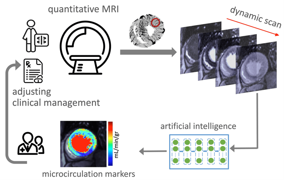
Microcirculation refers to the network of small vessels that supply oxygen-rich blood to the surrounding tissue. Patients with signs and symptoms of cardiovascular disease may suffer from microvascular disease, that is, the dysfunction of microcirculation in their heart muscle, which in turn, represents an important target for established or novel medical therapies. KCVRC scientists develop cutting-edge translational magnetic resonance imaging (MRI) techniques to investigate coronary microvascular function and to discover new markers that track the health of coronary microcirculation and quantify its response to therapy.

Human circulatory system showing heart anatomy

Improving the clinical paradigm for assessing endotypes of microvascular dysfunction

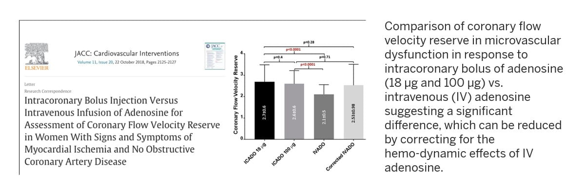
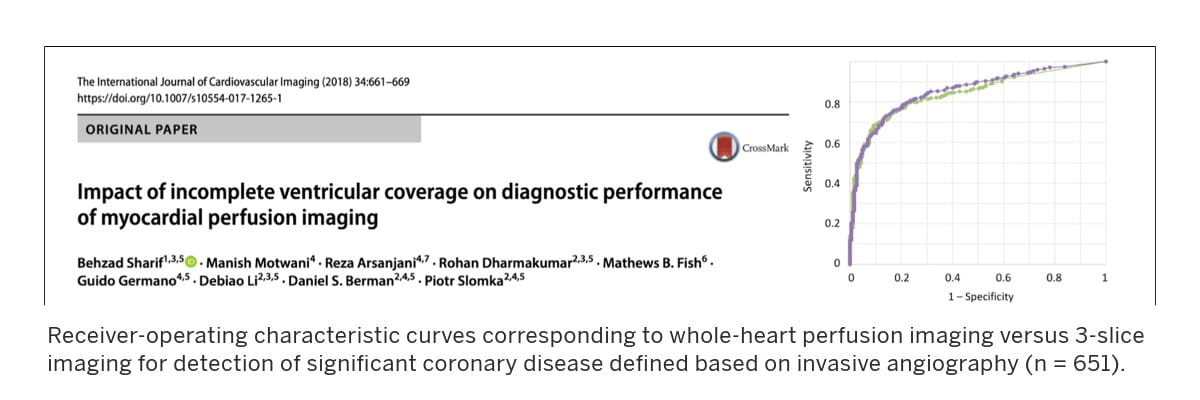
Led by Behzad Sharif, PhD, KCVRC investigators within the Microcirculaton Research program are advancing new approaches for noninvasive and minimally invasive assessment of patients with microvascular dysfunction by developing MRI-based techniques to quantify various markers of coronary microcirculation.

Two approaches advance the diagnosis and clinical management of patients affected by microvascular disease:

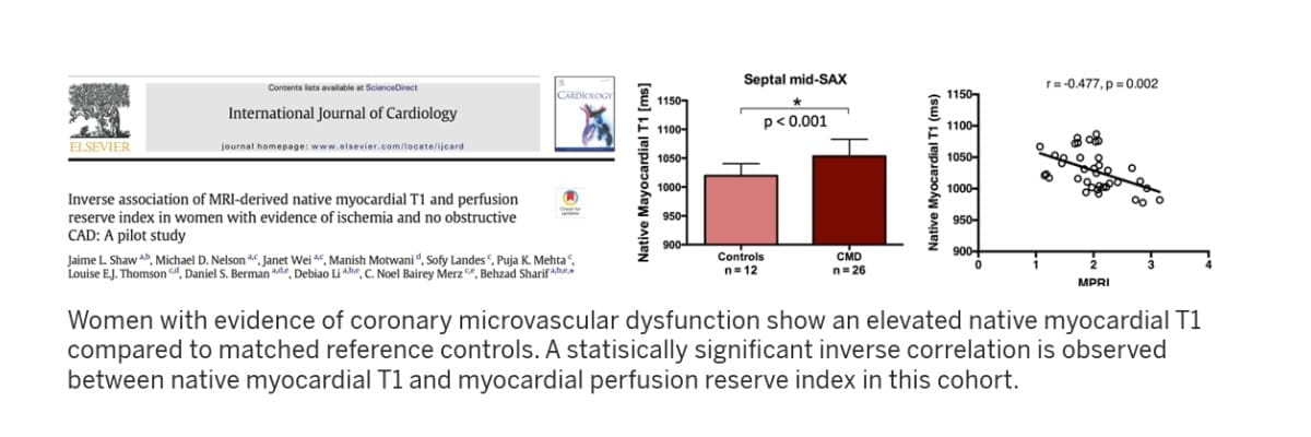
  • Reliable noninvasive assessment of microvascular health and function, including discovery and validation of new functional MRI markers of tissue microcirculation by leveraging multi-disciplinary innovations in magnetic resonance physics, computational engineering and machine learning
  • Development and validation of new interventional MRI approaches, enabling minimally invasive testing and endotype detection for microvascular disease, structural heart disease and cardiometabolic disorders.

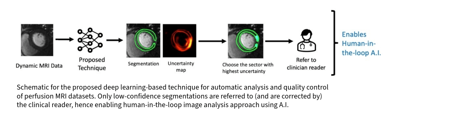
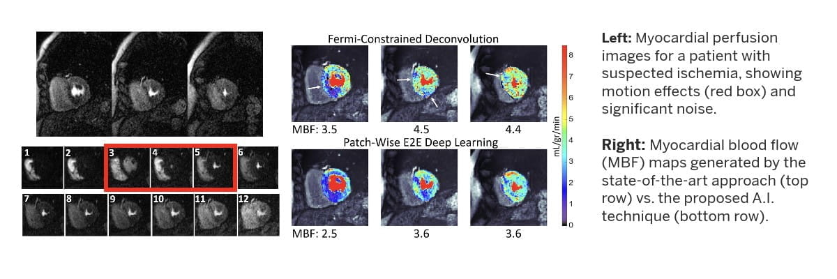
A significant component of our research effort is devoted to developing artificial intelligence-enabled MRI strategies (pulse sequences and accelerated image formation) and computational image analysis techniques aimed at enabling accurate characterization of the relationship between imaging markers of microcirculation and progression of disease, or response to therapy, in the context of nonobstructive coronary artery disease and cardiometabolic disorders.

Research in this lab is supported by the NIH National Heart, Lung and Blood Institute and the Lilly Endowment through the INCITE program.

This illustration shows the imaging-guided clinical management process for patients with coronary microcirculation dysfunction. The process begins with a quantitative MRI and then proceeds to the dynamic scan. Then, using artificial intelligence, microcirculation markers are identified and adjustments are made to clinical care based on the results.

The illustration shows the development process of image-guided clinical management of patients with dysfunction of coronary microcirculation. Once validated, these quantitative markers can be used by cardiologists to improve or adjust clinical management for patients who suffer from coronary microvascular dysfunction.


Active Funding

NIH R01 HL 153430 Noninvasive Testing of Coronary Microvascular Reactivity Using High-resolution Free-breathing MRI Disease

Funding Source: NHLBI, NIH   

PI: Behzad Sharif, PhD

Time Frame: 2020 – 2025

Description: Nearly half of the patients with chest pain who undergo invasive coronary angiography are found to have no obstructive or large-vessel coronary artery disease. The overall goal of this project is to develop MRI-based methods for diagnosis and monitoring of small-vessel (microvascular) coronary disease. Specifically, we will develop and translate a new noninvasive paradigm, powered by artificial intelligence, for clinical management of small-vessel coronary disease based on an MRI-based index of microvascular functional health and reactivity.

Indiana Collaborative Initiative for Talent Enrichment (INCITE) Program MRI-guided cardiac catheterization using minimally modified devices on a low-field scanner platform

Funding Source: Lilly Endowment, Inc.     

PI: Behzad Sharif, PhD

Time Frame: 2021 – 2026

Description: This project is an industry-academic collaborative effort between Dr. Sharif’s lab at KCVRC and MED Institute Inc. to evaluate the feasibility of MRI-guided cardiac catheterization using minimally modified devices on a commercially available low-field, ultra-wide bore MRI scanner platform. The long-term goal of this theme of research is to develop new methodologies for radiation-free interventional procedures aimed at accurate diagnosis of cardiovascular disease.


Completed Projects

NIH R00 HL 124323 Accurate Perfusion MRI for Improved Diagnosis of Microvascular Coronary Disease

Funding Source: NHLBI, NIH   

PI: Behzad Sharif, PhD

Time Frame: 2017 – 2021

Description: The goals of this project was to develop and test an artifact-free myocardial perfusion magnetic resonance imaging technique without the need for electrocardiogram triggering. 

Highlighted Publications

Temporal Uncertainty Localization to Enable Human-in-the-loop Analysis of Dynamic Contrast-enhanced Cardiac MRI Datasets

Yalcinkaya D, Youssef K, Heydari B, Simonetti OP, Dharmakumar R, Raman SV, Sharif B. Medical Image Computing and Computer-Assisted Intervention--MICCAI 2023. pp 453-462. doi: 10.1007/978-3-031-43898-1_44. 

This publication illustration shows a schematic for proposed deep learning-based technique for automatic analysis and quality control of perfusion MRI datasets.  From left to right: Dynamic MRI data, proposed technique, segmentation, uncertainty map, choose sector with highest uncertainty and refer to clinical reader. This enables Human-in-the-loop artificial intelligence.

A Patch-Wise Deep Learning Approach for Myocardial Blood Flow Quantification with Robustness to Noise and Nonrigid Motion

Youssef K, Heydari B, Zamudio Rivero L, Beaulieu T, Cheema K, Dharmakumar R, Sharif B. IEEE Proc Engineering in Med & Biol 2021:4051-4057. doi: 10.1109/EMBC46164.2021.9629630; PMID: 34892118.

on the left are myocardial perfusion images for a patient with suspected ischemia, showing motion effects and significant noise. On the right are myocardial blood flow maps generated by the state-of-the-art approach vs. the proposed AI technique

Inverse Association of MRI-derived Native Myocardial T1 and Perfusion Reserve in Women with Evidence of Coronary Microvascular Dysfunction and No Obstructive CAD: A Pilot Study

Shaw JL, Nelson M, Wei J, Motwani M, Mehta P, Thomson L, Berman D, Li D, Bairey Merz N, Sharif B. International J. Cardiology 2018; 270:48-53. doi: 10.1016/j.ijcard.2018.06.086; PMID: 30041981

publication details show that women with evidence of coronary microvascular dysfunction show an elevated native myocardial T1 compared to matched reference controls. A statistically significant inverse correlation is observed between native myocardial T1 and myocardial perfusion reserve index in this cohort.

Intra-coronary Bolus Injection versus Intravenous Infusion of Adenosine for Assessment of Coronary Flow Velocity Reserve in Women with Signs and Symptoms of Myocardial Ischemia and No Obstructive Coronary Artery Disease

Al-Badri A, Sharif B, Wei J, Samuels B, Azarbal B, Peterson JW, Anderson D, Gadh A, Henry T, Pepine CJ, Bairey Merz CN. J. American College of Cardiology (JACC): Cardiovascular Interventions 2018;11(20):2125-27; doi: 10.1016/j.jcin.2018.07.052; PMID: 30336817

publication highlight shows comparison of coronary flow velocity reserve in microvascular dysfunction in response to intracoronary bolus of adenosine vs. intravenous adenosine, suggesting a significant difference which can be reduced by correcting for the hemo-dynamic effects of IV adenosine.

Impact of Incomplete Ventricular Coverage on Diagnostic Performance of Myocardial Perfusion Imaging

Sharif B, Motwani M, Arsanjani R, Dharmakumar R, Fish M, Germano G, Li D, Berman DS, Slomka P.  Intern. J. Cardiovascular Imaging 2018;34(4):661–69. PMID: 29197024; doi: 10.1007/s10554-017-1265-1

publication highlight shows receiver-operating characteristics curves corresponding to whole-heart perfusion imaging vs. 3-slice imaging for detection of significant coronary disease defined based on invasive angiography
Principal Investigator
61027-Sharif, Behzad

Behzad Sharif, PhD, MSEE

Associate Professor of Medicine

Read Bio Behzad Sharif, PhD, MSEE

IU School of Medicine Collaborators

Gregory Anthony, PhD

Assistant Professor of Radiology & Imaging Sciences

Rohan Dharmakumar, PhD

Executive Director of the Krannert Cardiovascular Research Center

Kyle Frick, MD

Assistant Professor of Clinical Medicine

Ziad A. Jaradat, M.D.

Assistant Professor of Clinical Medicine

Rolf P. Kreutz, MD

Associate Professor of Clinical Medicine

Larry W. Markham, MD

Phillip Murray Professor of Pediatric Cardiology

Michael P. Murphy, MD

Cryptic Masons Medical Research Foundation Professor of Vascular Biology Research

Michael M. Ross, MD, MS

Associate Professor of Clinical Pediatrics

Research Team and Doctoral Trainees

Mehdi Amian, PhD student, Purdue University, Weldon School of Biomedical Engineering

Zhuoan Li, PhD student, Purdue University, Weldon School of Biomedical Engineering

Arian Mollajafari, PhD student, Purdue University, Weldon School of Biomedical Engineering

Mehmet Berk Sahin, PhD student, Purdue University, Elmore Family School of ECE

Hazar Benan Unal, MS, PhD candidate, UCLA, Department of Bioengineering

Dilek M. Yalcinkaya, MS, PhD candidate, Purdue University, Elmore Family School of ECE

Luis Zamudio, MS, Research Analyst, KCVRC

Shahriar Zeynali, MS, PhD candidate, Purdue University, Weldon School of Biomedical Engineering

Doctoral trainees pictured alongside Dr. Behzad Sharif. From left to right are: Arian Mollajafari Sohi, Mehdi Amian, Hazar Benan Unal, Dr. Sharif, Shahriar Zeynali, Dilek M. Yalcinkaya, Mehmet Berk Sahin and Zhuoan Li.

External Collaborators

East Coast

Michael Elliot, MD, Atrium Health, Charlotte, North Carolina

Jonathan Weinsaft, MD, Weill Cornell Medicine & NewYork-Presbyterian Hospital, New York


Midwest

Sean Chambers, PhD, Cook Advanced Technologies, West Lafayette, Ind.

Craig Goergen, PhD, Purdue University Weldon School of Biomedical Engineering, West Lafayette, Ind.

David Gross, PhD, MED Institute, West Lafayette, Ind.

Abolfazl Hashemi, PhD, Purdue Univ. Elmore School of Electrical & Computer Eng., West Lafayette, Ind.

Odayme Quesada, MD, The Christ Hospital, Cincinnati, Ohio

Bruno Tesini Roseguini, PhD, Purdue University Department of Health and Kinesiology, West Lafayette, Ind

Orlando P. Simonetti, PhD, The Ohio State University, Columbus, Ohio

Matthew Ward, PhD, Purdue University Weldon School of Biomedical Engineering, West Lafayette, Ind.

 
Southwest

Reza Arsanjani, MD, Mayo Clinic, Phoenix, Ariz.

Dipan J. Shah, MD, Houston Methodist & Weill Cornell Medical College, Houston, Texas


Southeast

Mohammad Al-Ani, MD, University of Florida College of Medicine, Gainesville, Florida

Robert M. Judd, PhD, Duke University School of Medicine, Durham, North Carolina

Puja Mehta, MD, Emory University School of Medicine, Atlanta, Georgia

Venkateshwar Polsani, MD, Piedmont Healthcare, Atlanta, Georgia


West Coast

C. Noel Bairey Merz, MD, Cedars-Sinai Smidt Heart Institute, Los Angeles, Calif.

Daniel S. Berman, MD, Cedars-Sinai Smidt Heart Institute, Los Angeles, Calif.

Janet Wei, MD, Cedars-Sinai Smidt Heart Institute, Los Angeles, Calif.


Canada

Bobak Heydari, MD, Libin Cardiovascular Institute, University of Calgary, Alberta, Canada

Andrew Howarth, MD, Libin Cardiovascular Institute, University of Calgary, Alberta, Canada