The Imaging Informatics programming team specializes in the exchange of health care data in real-time, leveraging HL7 and DICOM to create interventions in medicine. The team ingests data from radiology information systems, laboratory information systems, revenue cycle services, human resources, electronic medical records and specialty systems like point of care ultrasound to create information and knowledge for decision making. The core's clinical partners extend throughout Indiana, and its applications operate with several EMR, PACS and RIS vendors.
DORIS
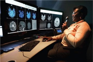
With an index of 20+ million radiology and pathology reports throughout the state, the Dig Our Radiology Information System (DORIS) superpowers our radiologists. In the clinical setting, DORIS allows clinicians to quickly find comparison studies, collect interesting cases for presentations and educate trainees. Researchers are able to quickly create cohorts for retrospective research and push the reports and images for de-identification. DORIS is connected to PowerBI, allowing data analysts at IU Health to generate performance dashboards.
Pipeline
Pipeline is the ‘pizza tracker’ of the radiology workflow. It is a web application designed to run at the nurses station in an emergency department, showing a list of all radiology exams for current patients. This list presents relevant information based on the exam status, changing phone numbers from modality technologist to radiologist as the exam is processed, displaying time stamps so clinical staff can follow up on exams that are delayed, and displaying the radiology report so they do not have to go into yet another system. Pipeline also functions for department radiologists, creating one worklist that collects data from the numerous EMR/RIS/PACS systems and allowing them to filter through most metadata for the exams they need to work on.
Ready access to relevant real-time information in medical imaging offers several potential benefits. Knowing both when important information will be available and that important information is available can facilitate optimization of workflow and management of time. Unexpected findings, as well as deficiencies in reporting and documentation, can be immediately managed. The Imaging Informatics core develops and has implemented a real-time web-centric dashboard system for radiologists, clinicians and support staff. The dashboards are driven by multi-sourced HL7 message streams that are monitored, analyzed, aggregated and transformed into multiple real-time displays to improve operations within our department. The framework is called Pipeline.
Ruby on Rails, JavaScript, HTML and SQL serve as the foundations of the Pipeline application. HL7 messages are processed in real-time by a Mirth interface engine which posts exam data into SQL. Users utilize web browsers to visit the Ruby on Rails based dashboards on any device connected to the hospital network. The dashboards will automatically refresh every 30 seconds using JavaScript. The Pipeline application has been well received by clinicians and radiologists.
RECIST Calculator
Response Evaluation Criteria in Solid Tumors (RECIST) 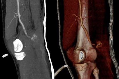 guidelines are utilized to objectively measure and analyze tumor treatment response in patients on clinical trials. RECIST requires radiologist measurement of solid tumors longitudinally through time. This presents a workflow issue, as varying radiologists may incorrectly identify tumor targets and may report the RECIST measurements differently. The Imaging Informatics core describes a web interface that simplifies RECIST measurement tracking, performs RECIST calculations and generates standardized report text. This system was implemented as a standard of care for all solid tumor patients, including those not on clinical trials, at the local children’s hospital. This project is called the RECIST Calculator (RC).
RECIST Measurements involve both radiology and oncology specialties. Both specialties use similar terminology to define conflicting concepts. For this paper, the Imaging Informatics core defines a ‘study’ as the set of patient encounters encompassing intervention or treatment for a given patient over a period of time; an ‘encounter’ as a series of images from one or more imaging modalities from which a single set of RECIST measurements are acquired by a radiologist; ‘image series’ as a set of images from an imaging modality.
For a given study, measurements are acquired longitudinally through time over many encounters and potentially varying modalities. The current workflow for RECIST measurements presented several opportunities for improvement at Indiana University School of Medicine, including:
- Tumor selection across encounters is not consistent. This is often due to the length between encounters. Additionally, due to the volume and nature of business, it is likely that any given encounter will be read by differing radiologists.
- There is no consistent RECIST reporting template. Specific measurements are often conveyed via paper forms.
- RECIST measurements are stored in oncology research databases, outside of the Electronic Medical Record (EMR). Measurements are manually entered from paper forms by staff.
- Clinically valuable RECIST calculations, such as percent change in tumor sizes (individual and overall) are not available for patients not enrolled in clinical trials that require RECIST measurements.
It was clear that IU School of Medicine would require a specialized tool to manage RECIST measurements. This tool would enable diverse radiologists to quickly identify prior measurement locations, standardize reported RECIST measurements and calculations and store RECIST measurements within the EMR; in such a way that would fit within the regular clinical workflow and be available to oncologists and radiologists to aid with patient care.
Stickr
STICKR merges radiology and pathology reports to allow for retrospective review. Radiologists receive notifications of reports to evaluate, marking their reports as concordant or discordant. This allows for retrospective self-evaluation and education, while generating metrics surrounding adequacy and diagnostic rates.
Research Study Tracking and JANUS
The Research Study Tracking tool transformed the Office of Research Imaging (ORI) project tracking from a manual and paper-based process to a fully digital workflow. This tool collects the information about the study and keeps everything up-to-date through interfaces with IRB and financial services. ORI can report on all the active studies, funding and staff members as needed for equipment grants, auditing and projecting staff workloads. Whether it’s the imaging protocol, radiopharmaceuticals, staff members or much more, every question about an imaging research activity can be answered through the study tracking tool.
JANUS is the gateway to ORI. This tool allows investigators and coordinators to submit new projects, schedule patients for scans and request other services that ORI provides. JANUS coordinates ORI staff work and updates are provided in real-time for investigators.
RIS for Research
The RIS for research has the functions you would expect from a RIS — it schedules patients, reserves equipment and collects dictations from voice recognition. The Imaging Informatics core's RIS also schedules radiopharmaceutical production at time of scheduling, ensuring patient schedulers can see the strict radiopharmaceutical timelines. This system is connected to Indiana University’s financial and the Research Study Tracking tools, automating billing and managing protocols. Future developments include a modality worklist function and DICOMWEB integration with PACS.