Body of Knowledge

Matthew Harris Apr 08, 2020
IT’S EARLY NOVEMBER, and more than 140 first-year students at Indiana University School of Medicine are quietly filing into a lab of the Medical Science Building in Indianapolis. Slowly, they tug on black nitrile gloves and cluster around 25 tables. Atop each table sits a steel container shaped like a casket. Flipping latches on the sides, they open the lids. Inside, resting on metal slabs before each student, they find a gift of the greatest intimacy, one that confronts them with the reality of their new vocation like nothing else: a human body. In life, these profound machines of skin, muscle and bone were animated by people. At some point, each person made an important choice that, upon death, their Earthly vessels would become visceral textbooks. For the 365 first-year students at the School of Medicine’s nine campuses, it’s a shared rite of passage on the way to becoming a physician. Months earlier, these students could barely glance at a body. Now, they know how to make long incisions, separate muscle and tendon, and trace the winding pathways that ferry blood and sensation to the tips of each limb. They have come to understand the body’s complex terrain. And today, these future healers will grasp the organ that, above all others, makes us truly human: the brain.
MUCH HAS CHANGED in the preparation of new doctors in recent years. Today’s medical students can watch most of their lectures online. They learn, early on, how to use an ultrasound device that fits in the palm of their hands. They practice procedures on mechanized mannequins that make breath sounds. Yet, for all that, one essential teaching tool has remained essentially the same for 500 years. And it binds physicians across generations.
The anatomy lab, where young medical students are confronted with the subject of their life’s work, is a classroom like no other. Mannequins offer hints at the geography. However, the latest technology and software can’t account for differences between bodies—muscles of varying sizes, organs withered by disease, or the unexpected course of a splenic artery.
Quickly, students realize that the vivid, colorful illustrations from their textbooks often bear little resemblance to the embalmed reality before them. Those images “look like 20-year-olds plucked from the Olympic ski team,” said Andrew Deane, PhD, who serves as course director for human anatomy at IU School of Medicine. Typically, the bodies the students explore in the lab belonged to the old and the sick.
After enough time in the lab, the students find that the scent of formaldehyde seeps into every fiber of their clothing, and even the pores of their skin. As such, Deane advises them not to wear clothes they like. While their subjects are cadavers, they are also the students’ first patients. And this is where they learn their first lessons in professionalism. Deane tells the students not to snap pictures for study at home—and not to bring in visitors. It all comes down to one final point, on which Deane is firm: respect the donors who bequeathed their remains. “Everybody here knows what that means,” he said, “and everybody knows when you’ve crossed the line.”

THE LONG JOURNEY to the brain began in August. At Table 21, the students were assigned the body of a 76-year-old man who died from congestive heart failure. At their first introduction, they encounter their subject as he lay face up. “This was a real person,” says student Dhruv Solanki. “You have this instinct to be super gentle.”
The course begins with the dissection of their subject’s back, which requires the student to turn him over. Once that’s accomplished, they face a new question: who will make the first incision?
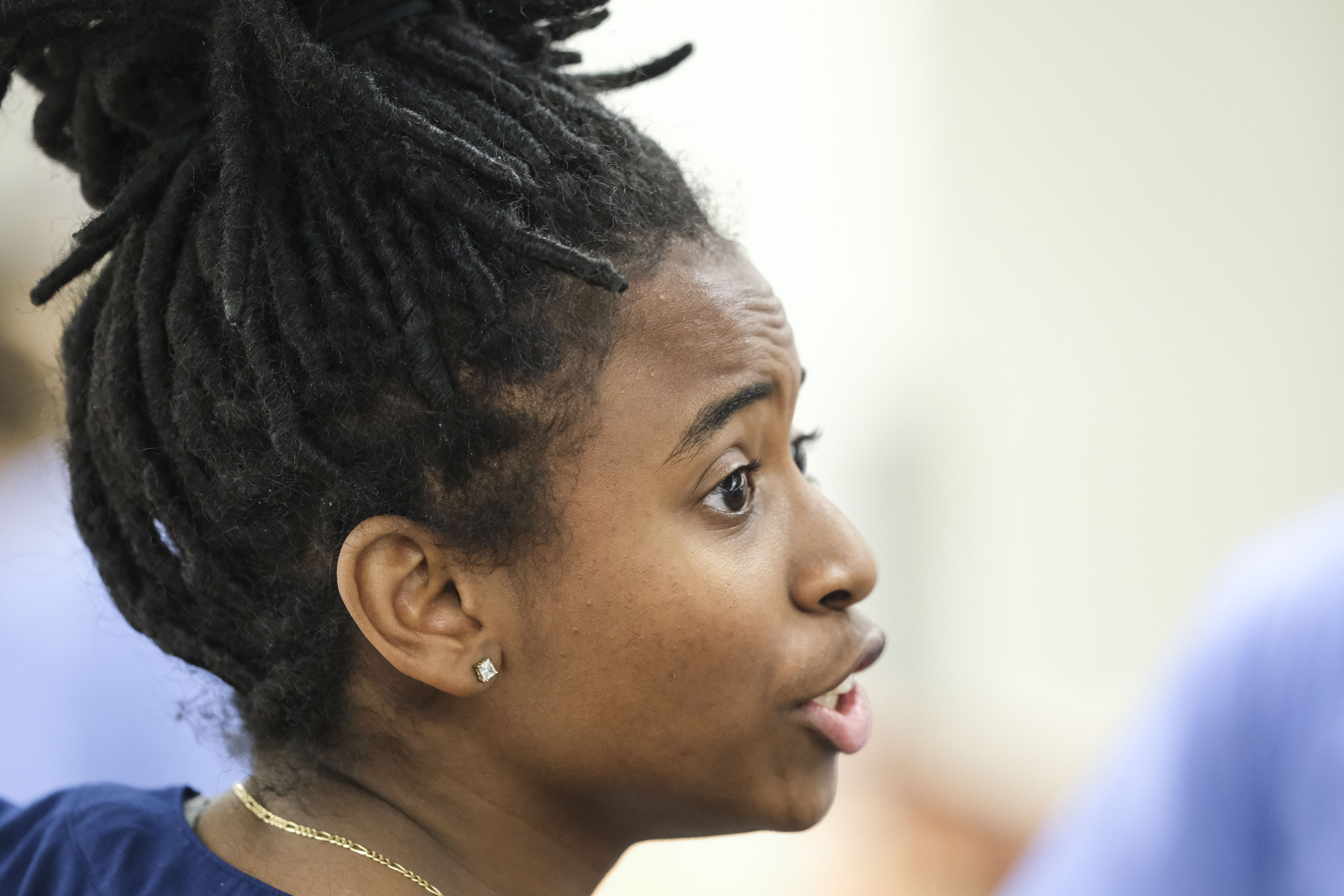
As she will throughout the course, Taylor Smith volunteers.
Inspired in middle school to be a surgeon after reading Ben Carson’s Gifted Hands, Smith, 25, is one of the few students who has previously taken a dissection-based anatomy course—a by-product of her Master of Science in Medicine from Mississippi College.
Along with being eager, Smith is apt to improvise. Gently, she uses a blade to trace a line down the middle of the back. “I’m really nervous,” she says.
Following her lead, Shivendra “Chevy” Singh makes another incision, just behind the earlobe, and continues his cut down the shoulder. Near the waist, Solanki takes over. He cuts horizontally, creating two flaps. At the start, the work is tediously slow—a two-hour lab isn’t enough time. But Deane is reassuring. He tells the students they will improve. “You will not be the same people anymore,” he says. “You won’t remember the problems you had on Day 1.”
With some coaching, the students reach their target—an exposed trapezius muscle. In this case, the muscle tissue is clearly delineated and easily identifiable. They slide fingers between muscles, with some measure of pride. “We have some great muscles,” Smith says.
OVER THE SEMESTER, the anxiety Solanki felt early on subsides. And the pace of the students’ work quickens. Other transformations follow.
Singh comes to marvel at the duality and contradictions of the human body: Its structures are well known, but the pictures in an atlas can’t account for variations between individuals. And then there is how it functions. “I didn’t realize how mechanical it all is,” Singh says. “It’s like, ‘How did this happen, and how did we discover it?’”
Few tasks convey this more than the difficult work of removing the spinal column to expose the cord it protects. When Singh casts a ponderous stare at the terrain, Smith asks, “You sure you want to do this?”
Singh, who injects a levity that counterbalances Smith’s focus, is undaunted. “This,” he says, “is my favorite part.”
At 26, Singh is the son of a radiologist and a researcher at the National Institutes of Health. His own path to IU was circuitous. He worked as a medical scribe and a cardiology researcher at the NIH. Singh was admitted to medical school in Australia, but he worried about the value of such a degree in the U.S. He applied to IU and, once offered a slot, came back stateside.
Medical school overwhelms some students, but not Singh. To get a jump on the week ahead, he watches old lectures on Saturdays. He comes into the lab without the others, preparing the patient for the next stage of the process. “He’s probably one of the most intelligent and culturally aware people I’ve ever met,” Smith says. “I don’t know where he wouldn’t fit in as a doctor. He could be whatever he wanted to be.”
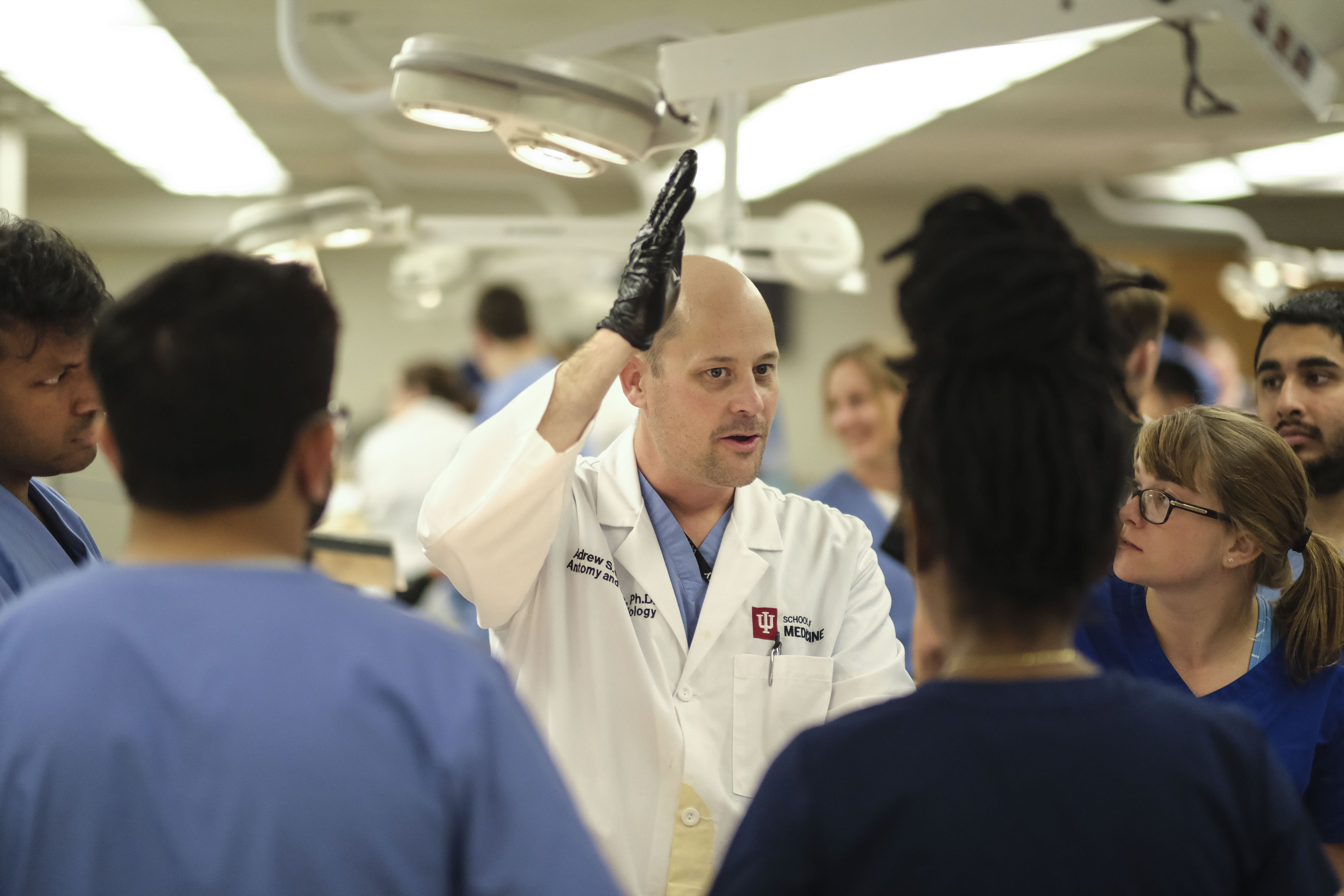
IN THIS LAB, when a student flags down Deane for a simple question, such as “Is this the MCL?” a lecture quickly breaks out.
Students from surrounding tables trickle over, and questions start coming from all directions. One prompts Deane to bend the leg of the man at Table 21, to point out various ligaments and to detail the evolution of the human knee.
Two decades ago, Deane stood where these students do now, experiencing the same mix of anxiety and hubris. Today, the skulls of ancient primates line the back shelves of his office, a collection amassed from fieldwork in paleoanthropology. At the University of Toronto, he learned anatomy.
For 15 years, including three at IU, Deane has taught the language of medicine to bright students aspiring to become doctors. To him, anatomy goes beyond learning the body’s geography. “They’re here to learn how to fix things,” he said. “If we just tell them where everything is, they won’t know why something is wrong.”
No matter where he looks, Deane sees high achievers in his classroom. Solanki is one of them. From the time he was a boy, Solanki, 22, was fascinated by hospitals. “There was always something going on,” Solanki says. “That always drew me in.” While an undergrad at IU—Northwest, Solanki worked as a scribe at Porter Regional Hospital in Valparaiso.
As a medical student, Solanki is most at ease watching lectures and reviewing flashcards in his apartment. In the lab, he’s not afraid to grab a scalpel, but he’s also content to let others do the cutting. “Taylor and Chevy know what they’re doing,” he says. “I don’t want to make a mistake.”
As has been the case for generations, the class, the content and the pace of anatomy class can be taxing. Success hinges on synthesizing reams of information. Practical exams ask students to apply their knowledge.
After the first exam, almost 40 students show up begging Deane for help. Many have just discovered that late-night cramming is no longer a strategy for success. Soon, there is an epidemic of what Deane describes as “imposter syndrome.” “They walk around thinking it was a mistake they got into medical school,” he says.
Among other things, the syllabus calls for students to clean out the plexus of nerves tucked under the armpit, navigate the abdominal cavity to trace a network of arteries feeding organs, and separate their body into three sections. Some students shudder at the tasks.
Others—including Singh, Smith and Solanki—are tightly focused. When studying together, they don’t delve into metaphysical discussions. “Those profound moments won’t come until we’re actually dealing with patients on the verge of death,” Smith says. “Right now, we aren’t trying to save someone.”
But they are grateful to their patient, a man who gave his body to science—and for what they’ve learned from him. They intend to show their appreciation by applying the lessons he has to offer.
A HEALTHY HEART, the students know from their textbook and lectures, is an efficient machine, one greedy for oxygenated blood, with valves that open and close in rhythm. Grant’s Dissector, their road map for exploring the human body, plainly outlines the steps they need to take to see the mechanics of a heartbeat.
But as she stands atop a stool, peering inside an open chest cavity, Smith is troubled by what she sees. The pericardium, a fluid-filled sac around the heart, is swollen. After some careful preparation, she and Singh lift the organ from the chest.
“Guys, this is not a good heart,” Smith declares.
“Could it have contributed to his death?” Singh asks.
“Definitely,” says assistant professor Elizabeth Augusto.
Later, a teaching assistant will lay out a possible pathology. Blood and fluid slowly filled the sac, compressing the organ. Over time, the heart’s strokes became shorter. The muscle of its walls thickened. The size of its chambers shrank in what became a bad feedback loop.
As they proceed with the dissection, Smith thinks she’s found a coronary sinus, a landmark that would tell them where they will need to cut to open heart chambers on the right side. They should also be able to easily find a groove that contains the left anterior descending artery. But their patient isn’t cooperating.
“I see no blood vessels at all,” Singh says.
The organ should be the size of a fist, but it looks small. Peeling back a flap and peering inside, the sight isn’t reassuring. “Oh my,” Singh says. “We’re missing stuff, and I’m not even sure why.”
The struggles distill for Singh a crucial lesson. The body is resilient and can withstand almost any acute shock. But the lives we lead and the choices we make—poor diet and lack of exercise—can leave it vulnerable. “There’s plenty of things that can go wrong without us,” Singh says. “But the only way we can truly hurt the body is if we don’t take care of it.”
THE BRAIN IS the pinnacle of this exploration of the human body. Once the difficult task of separating the skull is complete, the students can’t simply lift the organ out onto the table. They need their professor’s expert help.
Once he arrives, Deane carefully uses a foot-long scalpel to separate the brain from its connection to the brain stem. He asks a medical student to ball up a fist to represent the cerebellum, the major feature of the hindbrain.
Visible now are tiny veins that run across the surface of the brain. So are crimson-colored tributaries that once ferried blood to every fold. Near the front of the skull, students see the cable of optic nerves running to each eye.
Deane delicately removes the remaining bonds holding the brain in place.
As he goes about his work, Deane’s students hang back, silent and awestruck. “You wonder about all the thoughts that went through that head,” Singh says later.
It turns out you only need five minutes to free the brain from its resting place. And when he does, Deane smiles, turns, and gently places it in a pair of cupped hands. “Congratulations,” he says. “You have a six- to eight-pound brain.”
For a few fleeting moments, each student cradles the brain. They do not ponder its more profound significance. Their instructions aren’t a call to discuss what makes up our essence. Or to wonder about how our personality, memories and humanity are filed away in its folds and ridges.
For now, it’s enough that they can identify the Circle of Willis, a labyrinth of arteries that carry blood to the brain, or the sites where folds attach it to the skull. And just as quickly as the brain is delivered into their hands, a lab assistant arrives to collect it in a Ziploc bag.
Gingerly, the students place the brain inside and watch it disappear into another room, where it will be stored until the next semester. The next time they lay eyes on its folds may come during their class on neuroscience and behavior. “Whatever it has to teach,” Singh says, “will be for somebody else.”
To support medical education, contact Caitie Deranek Stewart at cderanek@iu.edu or 317-278-2133.
Matthew Harris
Matthew Harris is a communications specialist in the Office of Gift Development. Before joining the School of Medicine in 2015, he was a reporter at newspapers in Pennsylvania, Arkansas, and Louisiana. He currently lives in Indianapolis with his wife and two basset hounds.