In the breast imaging reading room at Indiana University Health Methodist Hospital, Stephanie Holz, MD, fellowship director for breast imaging in the Department of Radiology and Imaging Sciences at Indiana University School of Medicine, sat down to discuss the importance of mammography and the role it plays in the diagnosis of breast cancer.
What is breast imaging?
Breast imaging encompasses mammography, ultrasound, biopsy and breast magnetic resonance imaging (MRI). In this specialty, radiologists image the breast, biopsy it and, based on their findings, direct the patient to the appropriate care.
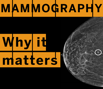
A mammogram of breast tissue where cancer was discovered (circled).
What is a mammogram?
A mammogram is an image taken of the breast tissue to determine early signs of breast cancer. For a mammogram, a patient stands in front of a specialized X-ray machine while their breast tissue is compressed. Through the process, the tissue is spread out and a series of images are captured. Mammography is unique in that it is one of the few screening tests that research has proven to reduce morbidity and mortality.
Are only those who identify as a woman at-risk for breast cancer?
No. Anyone who has breasts or an enlargement of breast tissue can be at-risk for breast cancer.
When should someone begin receiving mammograms?
The American College of Radiology and the Society of Breast Imaging recommends an annual mammography starting at age 40. An individual should consult with a physician if they think they could have a higher risk for breast cancer or notice anything unusual.
An ultrasound where breast cancer was discovered (gray mass) in the center.
What’s the difference between a screening mammogram and a diagnostic mammogram?
A screening mammogram is what patients receive during their regular annual check-up once they reach age 40. The majority of the patients we see receive this test. A diagnostic mammogram is taken to further examine suspicious changes in the breast (lumps, nipple discharge, skin changes or abnormalities observed from a chest scan), along with a clinical exam and ultrasound.
What’s the difference between bright and dark tissue?
When looking at a mammogram scan, the dark tissue is the fat and the bright tissue is the dense breast tissue. So if somebody has more than 50 percent of the white tissue, that’s called a dense breast. Locating the cancer amid a lot of bright tissue can be very challenging, like looking for a snowball in a snowstorm.
What are some new advances in breast imaging?
Within the last couple of years we’ve added automated breast ultrasound, which provides secondary imaging for individuals with dense breast tissue. This ultrasound has enabled physicians to locate cancers that would have otherwise gone undiagnosed. Another advancement in breast imaging is the abbreviated breast MRI protocol. Steven Westphal, MD and I are participating in this research that is looking at how abbreviated breast MRI performs versus mammography with tomosynthesis, a technique used to detect early stages of breast cancer. Once all the data has been collected on abbreviated breast MRI research, it could provide a new, less costly and more convenient option for certain individuals.
Annually, over 5,600 breast imaging screenings take place at IU Health Methodist alone, with screenings often increasing in October during Breast Cancer Awareness Month. Thousands of additional screenings are performed at IU Health locations throughout Indiana with IU School of Medicine radiologists playing critical roles in diagnosis and prevention.
Learn about breast cancer research at IU School of Medicine and check with an IU Health provider to learn more about what to expect from a mammography exam.
Breast cancer research IU Health Provider
