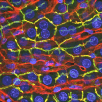
“Standard” confocal microscopy
For most applications, i.e., simple fluorescence imaging of fixed cells or tissues, most investigators will want to use the Olympus FV1000 or one of the Leica SP8 systems. These are user-friendly systems that support collection of multi-color, three-dimensional fluorescence images.
Learn about fluorescence and confocal microscopy
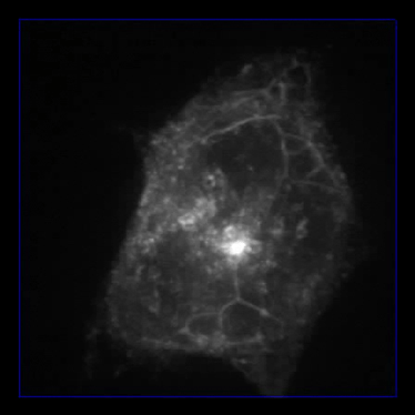
Microscopy of living cells
While any system can be used to image living cells, the Nikon Live Cell Imaging system supports the highest speed and lowest photodamage. The system is equipped with an epifluorescence path for the highest sensitivity and a spinning disk confocal path for high-speed, 3D imaging.
Learn about microscopy of living cells
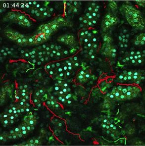
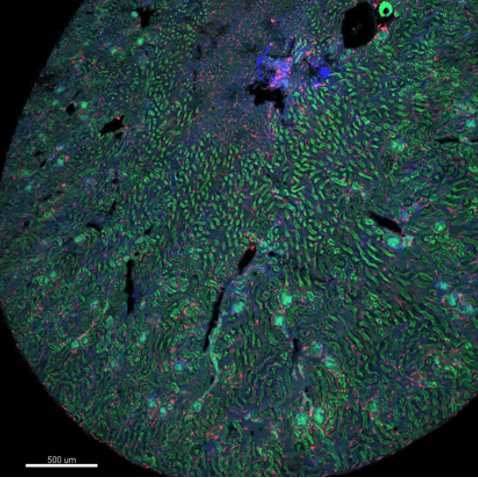
Multiphoton microscopy
Multiphoton microscopy is the technique of choice for imaging tissues in living animals. The three multiphoton systems and the fully-equipped surgical suite of the ICBM have been used for intravital microscopy of the kidney, liver, pancreas, bone marrow, lymph nodes, skin, mesentery and lung.
Learn about multiphoton-microscopy
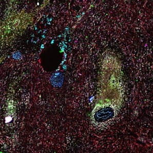
Fluorescence lifetime microscopy (FLIM)
A variety of biological properties can be assayed by measuring effects on the fluorescence lifetime of a biomolecule or probe. Studies utilizing FLIM, or the closely-related technique FCS, can be conducted using the ISS Alba system, or for studies in living animals, using the Leica SP8 DIVE system.
Learn about FLIM
Highly multiplexed CODEX microscopy
Multiplexed fluorescence microscopy of 40 or more probes can be conducted using CODEX fluorescence microscopy.
Learn about CODEX technology
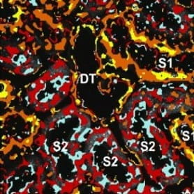
Volume visualization software
The ICBM has developed software tools for volume visualization and analysis.
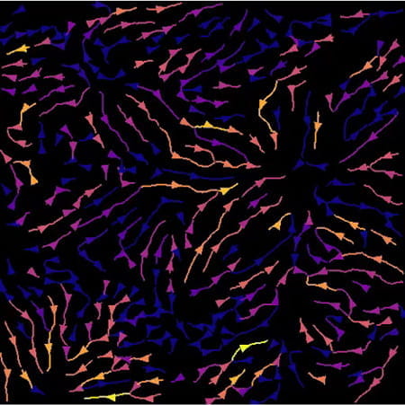
Digital image analysis software
In addition to equipping workstations with powerful commercial image analysis software such as Metamorph, Imaris, Halo and Akoya MAV, the ICBM has developed a variety of unique software tools over the past 20 years. Examples include software for volume rendering, motion artifact correction, measurement of microvascular flow, 3D segmentation and tissue cytometry.