2024 Speakers and Program

The online program is subject to change and final details may be distributed at the conference. Presenters, please contact Shari Parker (shupchur@iu.edu) to correct any outstanding errors.

Registration Details: 
Registered attendees can check in to the Breath Summit on Sunday, June 2 from 17:00 to 19:00, and on Monday from 07:30 to 09:00. The registration location in the Crowne Plaza Downtown Union Station will be in the foyer outside of the Illinois Street Ballroom on the second floor of the hotel.

Poster Session Details:
Posters should be hung at the beginning of the day and can be visited throughout the day during breaks. Poster presenters should attend their poster during their assigned poster session on Monday, Tuesday or Wednesday. Each poster session will be set up in the Crowne Plaza's Train Car North Platform, adjacent to the event's main conference room.

Please click within each session below to find additional details. 

International Congress for Breath Research

Sunday, June 2

17:00-19:00 — Hotel check-in, conference registration and a brief welcome reception with light hors d’oeuvres will take place in the Crowne Plaza Downtown Union Station. 

Monday, June 3

08:30-09:00 — Registration in the Crowne Plaza Downtown Union Station will be in the foyer outside of the Illinois Street Ballroom on the second floor of the hotel.

09:00-12:00 — Morning presentation session: 

“Breakthrough Developments and Discoveries”
09:00-09:10  Welcome presentation Mike Davis  
09:10-09:30 “From dynamic nature to human origin of breath isoprene - an investigative story”
Pritam Sukul
09:30-9:50
“Building a Metabolomic Database for Comprehensive Analysis of Exhaled Breath Composition and Biomarker Identification” Eva Borras
9:50-10:10  “Canine-Inspired Identification of Volatile Organic Compound Biomarkers for Noninvasive Health Monitoring: Opportunities in the 21st Century for Biotechnology”
Mark Woollam
10:10-10:30
Coffee break


10:30-11:30 — Mid-morning presentation session: “Advancements in Sampling”

10:30-10:50 “From research to routine: Meeting the evolving demands of clinical breath analysis with advancements in thermal desorption” Laura Miles 
10:50-11:10 “Assessment of exhaled breath VOC stability on TENAX® GR thermal desorption tubes”
Trenton Davis
11:10-11:30 “Addressing complexity: the evolution of breath biopsy and its potential in metabolic disease”
Matt Kerr

11:30-12:30 — Lunch buffet and exhibit hall session

12:30-14:30 — Afternoon presentation session: 

“Detecting Infections”
12:30-12:50  “Making an IMPACT: The results look promising for a P. aeruginosa breath test” Jane Hill
12:50-13:10  “Diagnosing Pulmonary Tuberculosis in Children through Breath Analysis: An Emerging Method” Ning Sonja Sun
13:10-13:30
“Selectivity to diagnose COVID-19 infection from exhaled breath volatiles (EBV) against other common upper respiratory infections” Mitchell McCartney
13:30-13:50
Coffee Break  
13:50-14:10
“Developing a Breath Test for Valley Fever using GC×GC Untargeted Volatilomics” Heather Bean
14:10-14:30
“Identifying putative volatile biomarkers for S. aureus methicillin-resistant and small-colony variant subtypes”  Daniela Gutierrez-Munoz
14:30-14:50  “Wearable Micro-GC device for non-invasive monitoring of sweat VOC patterns: advancements in disease diagnosis" Ruchi Sharma

14:50-15:00 — Break

15:00-15:30 — Rapid podium presentations (Each 2 minutes, one slide, 2 minutes for questions, timed)

“Assessment of exhaled breath VOC reproducibility on TENAX® GR thermal desorption tubes” Darakshan Zabin 
“ONELAB: applications of breath volatile analysis to rapidly screen for pathogenic infections”
Anesu Chawaguta
“Quality control framework for exhaled breath VOC analysis using thermal desorption”
Trenton Davis
“Quantitative analysis of a broad range of carbonyl compounds in exhaled breath for detection of COVID-19 during the periods of SARS-CoV-2 Alpha and Delta variants”
Xiao-An Fu
“Assessing response to phage therapy of M. abscessus using breath”
Antao Gao
“Human breath volatilome: a comparison of sampling and mass spectrometry methods for biomarker discovery in clinical research”
Elodie Lamy

15:30-15:50 — Special Presentation
Scott T. Dwyer will present, “The effect of wine on exhaled biomarkers and what to drink about it”

15:50-17:00 — Poster session

Volatilomic profiling reveals altered wild-type Pseudmonas aeruginosa metabolism compared to quorum sensing loss-of-function mutant strains  Waqar Ahmed 
Are we throwing the biomarkers out with the bath water? Scott Borden
ONELAB: applications of breath volatile analysis to rapidly screen for pathogenic infections
Anesu Chawaguta
QUALITY CONTROL FRAMEWORK FOR EXHALED BREATH VOC ANALYSIS USING THERMAL DESORPTION
Trenton Davis
Quantitative analysis of a broad range of carbonyl compounds in exhaled breath for detection of COVID-19 during the periods of SARS-CoV-2 Alpha and Delta variants
Xiao-An Fu
Assessing response to phage therapy of M. abscessus using breath
Antao Gao
Analysis of volatile aldehydes using secondary electrospray ionization mass spectrometry.
Stamatios Giannoukos
Determing volatile organic compound baselines in healthy breath with tin oxide sensors: a framework for non-invasive health monitoring.
Shivaum Heranjal
TD-GC-EI-PTR-MS for Exhaled Breath Analysis
Anne Jung
Micro-thermal Desorption Coupled to Gas Chromatography-Ion Mobility Spectrometry: Peppermint Protocol Standardization and Benchmarking
Kristian Kiland
Human breath volatilome: a comparison of sampling and mass spectrometry methods for biomarker discovery in clinical research
Elodie Lamy
A state-of-the-art laser technology for detecting breath molecules
Qizhong Liang
Defining VOC Signatures of Airway Epithelial Cells After Environmental Exposures
Angela Linderholm
Chip-Scale Mass Spectrometry for Point-of-Care Breath Diagnostics
Spiros Manolakos
Peptides and nanotubes - a machine learning and biological approach to VOC sensing
Oliver Nakano-Baker
Developing novel perovskite-based nanomaterials for the detection of volatile biomarkers in exhaled breath
Y Lan Pham
Interest of Soft Ionization by Chemical Reaction in Transfer (SICRIT) – high resolution mass spectrometry for exhaled breath analysis in clinical studies
Camille Roquencourt
Investigating Exhaled Volatile Organic Compounds in Healthy Breath Using Solid Phase Microextraction and Gas Chromatography-Mass Spectrometry
Eray Schulz
Extending Laser Absorption Spectroscopy towards Detection of Larger Volatile Organic Compounds in Breath
Miloš Selaković
ASSESSMENT OF EXHALED BREATH VOC REPRODUCIBILITY ON TENAX® GR THERMAL DESORPTION TUBES
Darakshan Zabin

17:00-17:30 — Anton Amman award: TBD

18:00-20:00 — Dinner will be provided and reception will occur in the Crowne Plaza Downtown Union Station.


Volatilomic profiling reveals altered wild-type Pseudmonas aeruginosa metabolism compared to quorum sensing loss-of-function mutant strains


Waqar Ahmed, Taoran Fu, Kamila Schmidt, Michael Brockhurst, Stephen J Fowler


University of Manchester, UK


Once a minimum cell density threshold is reached, Pseudomonas aeruginosa excretes small signaling compounds which activate functional genes responsible for virulence mechanisms. In this way, P. aeruginosa can persist in biofilms, form synergistic or parasitic interactions with commensal microbes, or invade a host causing life-threatening systemic infection1. In this pilot study we use gas chromatography-mass spectrometry to measure volatile metabolites from wild-type and loss-of-function mutant strains lacking in quorum sensing (QS) systems to uncover differences in cellular metabolism associated with QS signaling. Our initial results indicate 2-aminoacetophenone (2-AA) in culture headspace is lower in abundance for wild-type strains compared to lasR and rhl loss-of-function strains. This demonstrates that production of 2-AA, a metabolite of the PQS system is affected by the function of lasR and rhl and therefore confirms P. aeruginosa QS systems are interlinked and inter-dependent. This may have implications in breath biomarker discovery for infection2 as microbial metabolites have varied concentrations which are potentially dependent on intracellular interactions amongst other factors such as host response, nutrient availability, and cell concentration.


1Smith RS, Iglewski BH. P. aeruginosa quorum-sensing systems and virulence. Current Opinions in Microbiology 2003, 6(1), 56-60


2Ahmed WM, Fenn D, White IR, Dixon B, Nijsen TME, Knobel HH, Brinkman P, Van Oort PMP, Schultz MJ, Dark P, Goodacre R, Felton T, Bos LDJ, Fowler SJ, BreathDx Consortium. Microbial volatiles as diagnostic biomarkers of bacterial lung infection in mechanically ventilated patients. Clinical Infectious Diseases 2023, 76(6), 1059-1066.


The authors declare that they have no conflict of interest.


Developing a Breath Test for Valley Fever using GC×GC Untargeted Volatilomics


Emily A Higgins Keppler1,2, Heather L Mead3, Marley C Van Dyke4, Douglas F Lake1, D Mitch Magee5, Bridget M Barker6, Heather D Bean1,2


1School of Life Sciences, Arizona State University, Tempe, AZ

2Center for Fundamental and Applied Microbiomics, The Biodesign Institute, Tempe, AZ

3The Translational Genomics Research Institute (TGen), Phoenix and Flagstaff, AZ

4Microbiology Department, UT Southwestern Dallas, TX

5 Center for Personalized Diagnostics, Biodesign Institute, Arizona State University, Tempe, AZ

6 The Pathogen and Microbiome Institute, Northern Arizona University, Flagstaff, AZ


Background

Valley fever (coccidioidomycosis) is an endemic fungal pneumonia of the arid regions of North and South America. It is estimated there are 350,000 new cases of Valley fever each year, and in endemic and highly populated regions (e.g., Phoenix, Arizona and the San Joaquin Valley of California) up to 30% of community-acquired pneumonia (CAP) may be caused by Valley fever. The current diagnostics for Valley fever are severely lacking due to poor sensitivity and specificity, especially early in infection, which leads to delayed diagnosis, inappropriate treatment with antibiotics, lost productivity, and increased medical costs.

Objective

We are working toward the development of a breath test to discriminate Valley fever from other causes of CAP. In this study we performed untargeted volatilomics analyses of bronchoalveolar lavage fluid (BALF) samples from murine Coccidioides lung infections and BALF from persons with CAP to identify putative volatile biomarkers of Valley fever.


Methods

Murine model: All procedures were approved by the Institutional Animal Care and Use Committee (protocol 16–011) of Northern Arizona University. Three cohorts of mice were infected by intranasal inoculation with C. posadasii Silveira (n=6), C. immitis RS (n=6), or vehicle control (n = 4). After 10 days of infection, the mice were euthanized and 2 mL of BALF was collected for volatile metabolomics analyses and cytokine analysis by a mouse magnetic 26-Plex ProcartaPlexTM panel. Human samples: Mayo Clinic Arizona provided 55 BALF specimens, divided into three categories: coccidioidomycosis (n=14), non-Coccidioides CAP (n=29), and uninfected (n=12). All volatilomics samples were divided into technical triplicates and analyzed by headspace solid phase microextraction (SPME) and two-dimensional gas chromatography coupled with time-of-flight mass spectrometry (GC×GC-TOFMS). Random forest was performed on the human volatilomes to identify putative biomarkers to discriminate Valley fever from other causes of CAP.


Results

We detected 244 VOCs in the human BALF samples, eight of which could distinguish Coccidioides pneumonia from non-Cocci infected samples, and specifically from bacterial pneumonia, with > 95% accuracy. Data from the murine model suggest that a significant portion of the Valley fever volatiles are produced by the host and correlated with the immune response.


Conclusion

Combined, these pilot data indicate that a breath test to discriminate Valley fever from other causes of CAP is feasible and may facilitate antimicrobial stewardship through improved fungal detection and the stratification of disease severity.


Acknowledgements and Disclosures

This study was supported by an Arizona Biomedical Research Centre New Investigator Award to H.D.B. There are no conflicts of interest or financial disclosures for this study.


Are we throwing the biomarkers out with the bath water?


Scott Borden, Kristian Kiland, Crista Bartolomeu, Lucas Martins, Stephen Lam, Renelle Myers


Integrative Oncology, BC Cancer Research Centre, Vancouver, Canada


Background

Universal data pre-processing strategies that account for the influence of room air during breath biomarker discovery have yet to be established.  Meticulous care must be used to account for the VOCs in room air that a participant inhales prior to breath collection, as well as background exogenous environmental VOCs. Data preprocessing strategies to address this have consequently been aggressive to avoid false discovery, removing 100s of features. Accepted methods include requiring the breath signal to be larger than the room air signal or larger than the mean + 3× standard deviation. Other alternatives use a 3 µg/m3 threshold. Complicating this matter is the breath collection method used. Modern breath collection devices like the ReCIVAä differ significantly from one-breath exhalation devices as they involve the patient breathing in highly purified air for several breaths prior to starting, and throughout the collection. The sample is collected from an aggregate of multiple expirations over 5-10 minutes. Data processing has not been adapted for these devices.


Objective

To investigate the influence of exogenous environmental VOCs on breath samples acquired via the ReCIVAä device (breathing purified air).


Methods

Two healthy participants gave baseline breath samples, then were exposed to a selected VOC mixture for 30 minutes prior and during breath collection via ReCIVAä. Serial breath collections were then taken at 10, 20, 30, 60, and 120 minutes post exposure, in a clean room and compared for 12 target analytes. Peppermint, lemongrass, and cinnamon essential oils (EO) were prepared at 20 ppm in methanol and analyzed by TD-GC-MS to identify 12 target analytes. The EO mixture was dispersed in a small room (115 ft2) for one hour using a household diffuser prior to participant entering. All breath and room air samples were collected (1000 cm3) onto thermal desorption tubes and analyzed by thermal desorption gas chromatography mass spectrometry (TD-GC-MS).


Results

A total of 12 VOCs from the EOs were screened to assess the peak area response at each time point relative to the room air sample from the EO room. Room air samples from the EO room yielded large signals, with S/N values ranging from 2.8×10⁵ to 2.2×10⁶. Nine of twelve screened VOCs were not able to be detected in any ReCIVAä  breath data. The stark difference in signal is illustrated in Figure 1, demonstrating the room air peaks for neral, citral, and eugenol relative to the breath sample taken during EO exposure. Despite the large room air signals observed, nine of these twelve analytes were unable to be detected in ReCIVAä breath samples at any time point. Three of twelve analytes showed minor signals (<2.5% of signal relative to room air) in the exhaled breath sample taken in the EO room; these VOCs were those that arose from the peppermint EO and are often observed in breath.


Conclusions

Advances in breath sampling devices have greatly attenuated the influence of the environment on patient breath samples. Common preprocessing strategies currently employed may be overestimating the influence of room air and consequently removing relevant features.

Are we throwing the biomarkers out with the bath water? Graph depicting the cmparison of neral, citral, and eugenol peaks in room air, and from a ReCIVA  exhaled breath sample taken in the EO VOC-rich room. Additional time points are not shown for visual clarity, as they are also below detection limits

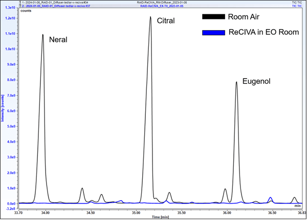
Figure 1. Comparison of neral, citral, and eugenol peaks in room air, and from a ReCIVAä  exhaled breath sample taken in the EO VOC-rich room. Additional time points are not shown for visual clarity, as they are also below detection limits


Building a Metabolomic Database for Comprehensive Analysis of Exhaled Breath Composition and Biomarker Identification


Eva Borras, Mitchell M McCartney, Cristina E. Davis


Mechanical and Aerospace Engineering, University of California, Davis, Davis, CA


Background

Exhaled breath condensate (EBC) is a matrix with particular interest for being a non-invasive and easily accessible biospecimen that can be collected without the need for complex procedures. EBC also contains valuable information of the composition of the respiratory system. Biomarker discovery and changes in metabolomic composition in EBC may indicate the presence of respiratory diseases such as asthma, chronic obstructive pulmonary disease (COPD), or even systemic conditions like diabetes.

While targeted approaches allow quantification of known compounds associated to specific diseases, they are focused on a predefined set of compounds. This can limit the coverage of the metabolome, overlooking unexpected metabolites that could be biologically relevant. Nevertheless, untargeted metabolomic approaches allow a broad and comprehensive profiling of the entire breath metabolome, enabling the discovery of novel biomarkers for respiratory or systemic conditions, and facilitating the exploration of entire metabolic pathways. The combination of both approaches can provide a better understanding of the metabolome, however, there are some challenges associated to untargeted liquid chromatography-mass spectrometry (LC-MS) metabolomics. One of the main limitations is the confidence in metabolite identifications, matrix effects, isomeric co-elutions or quantification of the new biomarkers, needing spectral databases or authentic standards for verifications.


Moreover, in the case of EBC, the sample amount is limited and often requires intense pre-concentration steps to detect metabolites. Analytical quality control practices are usually challenging when working with this matrix, and the re-analyzing of samples for a proper characterization (e.g. MSn) is often not possible. The use of authentic standards can enhance the analytical method, use as quality control, serve as reference compounds for validation, and index retention times by creating spectral libraries by commercial standards databases. These libraries contain mass spectra and retention time information for a wide range of metabolites that can be compared with experimental spectra from samples and allow better putative identifications. Additionally, these compounds can be quantified and asses concentration changes in EBC.


In this study, we present the development of a metabolomic database using commercially available metabolite libraries to enhance our understanding of the EBC composition and the identification of potential new biomarkers.


Methods

We compiled a metabolomic database by integrating four commercially available metabolite libraries, ensuring coverage across a wide range of metabolite classes. This comprehensive database, containing over 1500 metabolites, serves as a reference library for the identification and quantification of metabolites present over 400 EBC samples from different studies. LC-MS was employed for high-throughput profiling of EBC samples, with subsequent data processing and annotation using the established metabolomic database.


Results

The new metabolomic database includes a diverse array of known metabolites, including volatile organic compounds, lipids, amino acids, and small molecules from 400 samples. Analysis of EBC samples from different cohorts revealed a complex and dynamic metabolomic profile. Preliminary findings indicate significant variations in the abundance of specific metabolites associated with asthma, influenza, COVID-19, or even effects of flu vaccination of exposure of wildfires. These results can contribute to our better understanding of metabolic pathways implicated in respiratory and systemic health.

ONELAB: applications of breath volatile analysis to rapidly screen for pathogenic infections


Anesu Chawaguta1, Paul Brinkman2, Zoltan Gyongyi3, Chris A. Mayhew1, Veronika Ruzsanyi1, Daniel Sanders4, Makoto Sawano5, Matthias Schlögl6 and C. L. Paul Thomas7


1Institute for Breath Research, Universität Innsbruck, Innsbruck, Austria

2Department of Respiratory Medicine, Amsterdam UMC, University of Amsterdam, Amsterdam, Netherlands

3University of Pec, Pecs, Hungary

4G.A.S. Gesellschaft für Analytichesensensorsysteme GmbH, Dortmund, Germany

5Saitama Medical University, Saitama, Japan

6Solgeium OG, Linz, Austria

7Bioxhale Ltd, Leicester, United Kingdom


Background

The outbreak of the COVID-19 pandemic showed how unprepared and uncoordinated governments were to the entry of a novel virus into the human population. A major problem was the lack of a rapid response to mitigate the impact of the SARS-CoV-2 virus on society. To help lessen the potential impact of future emergent viral infections, a recently funded Horizon Europe Programme and UK Research and Innovation project called “ONELAB” is developing next-generation modular rapid response mobile laboratories for flexible, scalable and multi scenario deployments of analytical devices and clinical tests to provide point-of-need tests to determine infections with a high-level of situational awareness [1].


Objective

A key objective of ONELAB is to screen people rapidly and accurately for pathogenic infection at the outbreak of a disease when much is still undefined. An aim is to provide rapid triage to determine a person’s infection status. A key element to this diagnostic capability is the use of acute volatile infection biomarkers present in exhaled breath that can be used to determine whether a contagion is present or not. A proof-of-principle study has already demonstrated the potential of using breath volatiles to diagnose SARS-CoV-2 infection [2]. ONELAB significantly expands on this earlier seminal work by undertaking longitudinal observational epidemiological studies over two winters to identify and monitor volatiles in exhaled breath that are associated with acute respiratory tract infections, involving four clinical partners based in Europe and Japan. Using Gas Chromatography Mass Spectrometry (GC-MS) for volatile discovery and GC-Ion Mobility Spectrometry (GC-IMS) for near-to-real time breath pattern profiling, we will acquire the knowledge to develop GC-IMS as a technological platform for use in a non-invasive breath infection-screening breath test as a first line of defence procedure to protect people, healthcare systems, and economies.


Discussion

In January 2024, ONELAB started to obtain breath samples for analysis from infected patients and from controls from all four clinics, with a target of 1000 samples to be collected and analysed, making this the largest breath pathogenic infection study to date. The GC-IMS pathogenic volatile breath fingerprints are being incorporated into innovative machine-learning pattern recognition algorithms to profile the signature patterns of infection in the breath volatilome for direct use in screening. Preliminary GC-MS and GC-IMS breath sample analyses will be presented together with our initial outcomes from the machine learning algorithms.


Acknowledgement

We acknowledge the EU HORIZON Innovation Actions HORIZON-CL3-2021-DRS-01-05, Project Number 101073924 (ONELAB) for funding this work.


References

[1]    https://onelab-project.eu/

[2]    D. Ruszkiewicz et al eClinicalMedicine 29-30 (2020) doi: 100609 doi: 10.1016/j.eclinm. 2020.100609


Assessment of Exhaled Breath VOC Stability on Tenax® GR Thermal Desorption Tubes


Trenton J. Davis1,2, Ivan V. Ivanov3, Darakshan Zabin1, Heather D. Bean1,2


1School of Life Sciences, Arizona State University, Tempe, AZ

2Center for Fundamental and Applied Microbiomics, The Biodesign Institute, Tempe, AZ

3Department of Veterinary Physiology & Pharmacology, Texas A&M University, College Station, TX


Background

The analysis of exhaled breath volatile organic compounds (VOCs) is increasingly being leveraged to study human health and disease including respiratory infections, asthma, and metabolic disorders. Sampling of breath VOCs often involves transferring breath samples onto sorbent-packed thermal desorption tubes (TDTs) for concentration, transport, and storage. Due to constraints such as distant sampling locations and instrument capacity, samples cannot always be same-day analyzed and must be stored for a future processing date, and sometimes must be shipped using air or ground transportation. This brings forth questions regarding the most appropriate storage method, specifically with regards to storage length, for maintaining sample integrity.


Objective/Aims

The goal of this study was to examine the effects of storage time at 4 °C and shipping method (air vs. ground shipping) on the recovery of exhaled breath VOCs from Tenax® GR TDTs.


Methods

All procedures were approved by the U.S. Army Medical Research and Development Command (USAMRDC), Office of Human and Animal Research Oversight (OHARO), and Office of Human Research Oversight (OHRO; log numbers E03915.1a and E03915.1a-1). Three experiments were conducted: Experiment 1 tested the variable of storage time alone; Experiment 2 tested storage time plus air shipping vs. no shipping; and Experiment 3 tested storage time plus ground shipping vs. air shipping vs. no shipping. For each experiment, breath was collected in 10 L Tedlar bags from four subjects and split onto 12 to 14 technical replicate Tenax® GR TDTs. Exp 1 samples were stored at 4 °C and technical duplicates were analyzed using thermal desorption – comprehensive two-dimensional gas chromatography – time of flight mass spectrometry (TD–GC´GC–TOFMS) after 0.5, 1, 2, 4, 8, 16, and 32 days; Exp 2 technical duplicates were analyzed after 8, 16, and 32 days; Exp 3 technical duplicates were analyzed after 16 and 32 days.


Results

We found no significant effect of storage time for up to 32 days on the relative abundances and the numbers of detected VOCs, or on the reproducibility of the breath VOCs on technical duplicate samples. Similar results were observed when air and ground shipping were taken into consideration.


Conclusions

Overall, these results demonstrate that exhaled breath VOCs collected and shipped under these conditions can be stored for upwards of a month without jeopardizing sample integrity.


Acknowledgements and Disclosures

This work was supported by the Defense Advanced Research Projects Agency (DARPA) Fatigue Assessment via Breath (FAB) study (Cooperative Agreement HR00112220040; Roozbeh Jafari and Linda Katehi, PIs). The views, opinions, and/or findings contained in this material are those of the authors and should not be interpreted as representing the official views or policies of the Department of Defense or the U.S. Government. No official endorsement should be inferred. There are no conflicts of interest or financial disclosures for this study. 


Quality Control Framework for Exhaled Breath VOC Analaysis Using Thermal Desorption


Trenton J. Davis1,2, Bilal N. Ali1, Darakshan Zabin1, Heather D. Bean1,2


1School of Life Sciences, Arizona State University, Tempe, AZ

2Center for Fundamental and Applied Microbiomics, The Biodesign Institute, Tempe, AZ


Background

Sampling exhaled breath volatile organic compounds (VOCs) for offline analysis often involves multiple steps, including the collection of exhaled breath in bags, transferring the breath via a pump to sorbent-packed thermal desorption tubes (TDTs) onto which the VOCs adsorb, storing and transporting samples to the analytical lab, and then desorbing the VOCs from the TDTs into the inlet of a gas chromatograph. Due to the complexities and expense of human sampling, exhaled breath samples are precious, and great care is undertaken to maintain and assess their integrity through the sampling and storage/handling process. These efforts are not always followed through to analysis, however, and many researchers fail to adequately evaluate analysis quality (e.g., sufficient sample desorption and injection). Taking from process engineering, we present an easily implemented statistical quality control framework for post-hoc monitoring of breath sample analysis via thermal desorption – gas chromatography (TD–GC).


Methods

All procedures were approved by the U.S. Army Medical Research and Development Command (USAMRDC), Office of Human and Animal Research Oversight (OHARO), and Office of Human Research Oversight (OHRO; log numbers E03915.1a and E03915.1a-1). Four subjects were recruited to provide three breath samples in 10 L Tedlar bags. All breath samples were collected on the same day, and within 120 minutes of sample collection, each sample was split into 0.6 L technical replicates by transferring onto Tenax® GR TDTs. TDTs (n = 152) were stored at 4 °C and technical duplicate TDTs were analyzed after 0.5 to 32 days of storage using thermal desorption – comprehensive two-dimensional gas chromatography – time of flight mass spectrometry (TD–GC´GC–TOFMS). Multivariate T2 single control charts of five low-variance VOCs detected across all samples were calculated using the R package “qcc”. Out-of-control (OOC) samples were defined as those with T2 statistics exceeding (1–0.0027)p, where p is the number of variables and 0.0027 is the probability of a Type I error for a single Shewhart chart at the mean ± 3 (s.d.).


Results

Using the multivariate Hoteling’s T2 statistic of five endogenous breath VOCs, we identified OOC breath samples at a rate not greater than 5% in an analysis of 152 breath TDT samples.


Conclusions

We show that this quality control framework can quickly identify OOC breath samples post-analysis, enabling the exclusion of the samples from downstream data processing, or the replacement of the OOC sample with a back-up technical replicate in order to preserve the sample size of the study.


Acknowledgements and Disclosures

This work was supported by the Defense Advanced Research Projects Agency (DARPA) Fatigue Assessment via Breath (FAB) study (Cooperative Agreement HR00112220040; Roozbeh Jafari and Linda Katehi, PIs). The views, opinions, and/or findings contained in this material are those of the authors and should not be interpreted as representing the official views or policies of the Department of Defense or the U.S. Government. No official endorsement should be inferred. There are no conflicts of interest or financial disclosures for this study.


Quantitative analysis of a broad range of carbonyl compounds in exhaled breath for detection of COVID-19 during the periods of SARS-CoV-2 Alpha and Delta variants


Zhenzhen Xie; James Morris1; Jianmin Pan; Elizabeth A. Cooke; Saurin R. Sutaria; Dawn Balcom; Subathra Marimuthu; Leslie W. Parrish; Holly Aliesky;Justin J. Huang; Shesh N. Rai; Forest W. Arnold; Jiapeng Huang; Michael Nantz; Xiao-an Fu


University of Louisville


Quantitative analysis of a broad range of carbonyl compounds in exhaled breath for detection of COVID-19 during the periods of SARS-CoV-2 Alpha and Delta variants Background: COVID-19 has caused a worldwide pandemic, creating an urgent need for early detection methods. Breath analysis has shown great potential as a non-invasive and rapid means for COVID-19 detection. Volatile organic compounds (VOCs) are generated by host responses through a series of lipid degradations including ketosis and inflammatory processes present in the lungs. Breath analysis techniques also demonstrated the potential for differentiation of variants of SARS-Cov-2. Many variants of SARS-Cov-2 create challenges for its detection and curbing the disease.


Objective

The objective of this study is to detect COVID-19, differentiate of Alpha from the Delta variant, and detect asymptomatic COVID-19 infection by analysis of carbonyl compounds in exhaled breath using a microreactor approach.


Methods

The study included a cohort of COVID-19 positive and negative subjects confirmed by reverse transcriptase polymerase chain reaction between March and December 2021. Carbonyl compounds in exhaled breath were captured using a microfabricated silicon microreactor and analyzed by ultra-high performance liquid chromatography-mass spectrometry (UHPLC-MS). A total of 341 subjects were enrolled in this study. Of these, 141 (85 males, 60.3%) (mean±SD age: 51.6±15.1 years) were COVID-19 positive and 180 (90 males, 50%) (mean±SD age: 45.2±14.9 years) were negative. Panels of ketones and aldehydes in breath samples were identified for detection of COVID-19 positive patients. Logistic regression models were employed for the data. The model performance was evaluated by the ROC curve with AUC, accuracy, sensitivity and specificity.


Results

Logistic regression models indicated high accuracy/sensitivity/specificity for alpha variant (98.4%/96.4%/100%), for delta variant (88.3%/93.0%/84.6%), for all COVID-19 positive patients (94.7%/90.1%/98.3%), and for asymptomatic patients (95.3%/71.0%/99.5%).


Conclusion

COVID-19 positive patients can be detected by analysis of carbonyl compounds in exhaled breath by microreactor approach. The technology for analysis of carbonyl compounds in exhaled breath has great potential for rapid screening and detection of COVID-19 and for other infectious respiratory diseases in the future endemics.

Assessing response to phage therapy of M. abscessus using breath


Antao Gao1, Jerry A. Nick2,3, Rebekah M. Dedrick4, Graham F. Hatfull4, Katie Poch2, Silvia Caceres2, Ahmad Mani1, Jane E. Hill1


1Department of Chemical and Biological Engineering, University of British Columbia, Vancouver, BC V6T 1Z4, CA

2Department of Medicine, National Jewish Health, Denver, CO 80206, USA

3Department of Medicine, University of Colorado School of Medicine, Aurora, CO 80045, USA

4Department of Biological Sciences, University of Pittsburgh, Pittsburgh, PA 15260, USA


Background

Mycobacterium abscessus (M.abs) is a nontuberculous mycobacteria (NTM) that may cause severe lung infection in people with cystic fibrosis (pwCF). High levels of intrinsic and acquired antibiotic resistance in M. abs result in limited treatment options and often lead to multiple relapses and low cure rates. This may prevent pwCF from receiving lung transplant. Phages are viruses that attack bacteria selectively, and their therapeutic use has been evaluated for difficult-to-treat infections, with recent successful phage treatments of M. abs in pwCF. The sensitivity of NTM microbial culture in cystic fibrosis airways can be as low as 30%. Thus, there is a growing need for better culture-independent markers of treatment response. Exhaled breath, capturing volatile compounds emitted by bacteria or the host, is a promising candidate for such biomarkers.


Hypotheses

1) Volatile biomarkers in breath can differentiate between M. abs positive and M. abs negative cases; and 2) Phage therapy for M. abs can be reflected in the variation of the abundance of volatile biomarkers in breath.


Methods

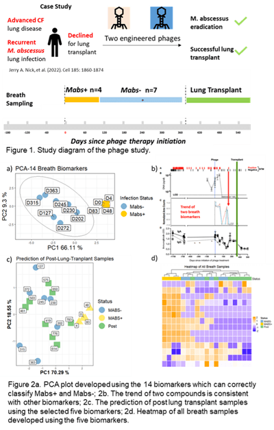
Two engineered phages were used to treat M. abs infection in an individual with advanced cystic fibrosis (CF) lung disease. A variety of markers combined with extensive cultures indicated eradication of M. abs following a year of treatment, allowing for successful lung transplant. Breath samples (3 Liter) were collected at multiple time points prior to the phage therapy till after the lung transplant operation (Figure 1). Breath samples were collected into Tedlar bags and concentrated onto thermal desorption tubes (TDTs) which were then analyzed using two-dimensional gas chromatography coupled with a time-of-flight mass spectrometry (GC×GC ToFMS). Chemical compounds were identified by spectral library match, and putative diagnostic features were selected by statistical analysis using R programming.

Results: Fourteen volatile compounds in breath were identified that differentiated between M. abs positive (M. abs+, n=7) and M. abs negative (M. abs-, n=4) breath samples (Figure 2a). The changes in the relative abundances of a subset of these 14 volatile compounds were consistent with variations in other biomarkers, including microbial culture, immunoglobulin (IgA and IgG), urine lipoarabinomannan (LAM), and M.abs DNA (Figure 2b). Five out of the 14 volatile compounds were detected in breath samples (n=8) collected after lung transplant, and these five biomarkers correctly classified all the breath samples as M. abs-, which conformed to the culture results (Figure 2c and 2d). Chemical identity of three compounds were confirmed by authentic chemical standards.


Conclusion

There are 14 compounds in breath of this patient that could diagnose M. abs lung infection and correlate to phage treatment efficacy during an M.abs lung infection in a pwCF.

The study protocol was approved by the University of British Columbia Research Ethics Boards. This study was supported by the US National Institutes of Health, R01 HL146228-01. The authors declare no conflict of interest.

graphics from the Breath Summit 2024 abstract, "Assessing response to phage therapy of M. abscessus using breath." Figure 1 depicts a study diagram of the phage study and several biomarkers.
 

Analysis of volatile aldehydes using secondary electrospray ionization mass spectrometry.

S. Giannoukos1, E. Lattouf 1, M. Emery1, R. Zenobi1

1 Department of Chemistry and Applied Biosciences, ETHZ, Zurich, Switzerland


Background

Aliphatic straight-chain aldehydes have been consistently identified in the breath of individuals with lung diseases through multiple detection methods, including mass spectrometry, ion mobility spectrometry, and electrochemical sensors. Research has revealed elevated levels of these exhaled aldehydes in patients with various lung conditions, including lung cancer, inflammatory and infectious lung diseases, as well as mechanical lung injuries. Quantifying straight-chain aliphatic aldehydes in the clinical setting is thus highly important.


Objective/Aims

This work focuses on the qualitative detection and quantification of volatile straight-chain aliphatic aldehydes using an online secondary electrospray ionization source coupled to a high-resolution mass spectrometry system.


Methods

A secondary electrospray ionization (SESI) source coupled to a high-resolution mass spectrometry (HRMS) system was employed for the first time to detect and monitor, both qualitatively and quantitatively, selected volatile straight-chain aliphatic aldehydes in the gaseous phase.  The generation of gas standards was done using a built-in-house vapor generator based on the controlled evaporation of volatile or semi-volatile chemical analytes and their diffusion into a carrier gas stream. The main components of the vapor generator are a) a mixing chamber, b) three individual temperature-controlled evaporation chambers in which liquid analytes are introduced through a side injection port, c) four mass flow controllers, and d) an automation platform controlled by software. SESI-HRMS is a powerful, well-established, and robust analytical technique ideal for in-depth breath metabolomics characterization, offering high sensitivity (low limits of detection), selectivity, fast (within seconds), and accurate analysis.


Results

Experiments were undertaken for propanal, butanal, pentanal, hexanal, heptanal, octanal, nonanal, and decanal at different concentrations and flow rates. Both individual compounds and mixtures were tested. Gas-phase experiments were performed at concentration levels from low ppt to low ppm and in both dry and humid conditions. The experimental results obtained showed a precise and repeatable production of gas standards with excellent linearity within the examined concentration range, low ppt detection limits, and fast response times.


Conclusions

For the first time, the qualitative and quantitative analysis of straight-chain aliphatic aldehydes was demonstrated using a dynamic vapor generator, capable of producing gaseous standards in a precise and reproducible way, with a SESI source coupled to a high-resolution mass spectrometer. The analytical performance of this approach was investigated by addressing essential analytical criteria such as limits of detection, limits of quantification of individual compounds, and the response in multi-component mixtures under dry and humid conditions in the full mass range m/z from 50 to 500.


Identifying putative volatile biomarkers for S. aureus methicillin-resistant and small-colony variant subtypes

Daniela F. Gutiérrez-Muñoz1,2, Daniel J. Wolter,3 Lucas R. Hoffman,3 Brandie D. Wagner,4 Heather D. Bean1,2

1School of Life Sciences, Arizona State University, Tempe, AZ

2Center for Fundamental and Applied Microbiomics, The Biodesign Institute, Tempe, AZ

3Department of Pediatrics, University of Washington, Seattle, WA

4Biostatistics & Informatics, Colorado School of Public Health, University of Colorado, Aurora, CO


Background

Seventy percent of persons with cystic fibrosis (pwCF) in the US are respiratory culture positive for Staphylococcus aureus, and this rate is climbing (1). With increasing S. aureus infections, the detection of clinically-relevant subtypes, such as methicillin-resistant S. aureus (MRSA) and small-colony variants (SCVs), is also increasing. Both subtypes, but especially thymidine-dependent SCVs, have been associated with worse patient outcomes (2, 3). Due to the high rates and co-occurrence of SCVs and MRSA (2), detecting and tracking the emergence of each of these S. aureus subtypes in CF lung infections is important for understanding the progression of CF lung disease and for formulating effective treatment options.


Objective/Aims

We are working to develop breath tests for detecting S. aureus MRSA and SCV lung infection subtypes in situ. As a first step in this process, we have characterized the in vitro volatile organic compounds (VOCs) produced by 110 S. aureus isolates from CF lung infections, representing four classes: methicillin-sensitive S. aureus normal colony variants (MSSA-NCVs), MSSA-SCVs, MRSA-NCVs and MRSA-SCVs.


Methods

The S. aureus clinical isolates in this study originated from pwCF and were phenotyped for methicillin sensitivity and SCV thymidine auxotrophy. The S. aureus cultures were resuspended from glycerol stocks in 100 ml LB-Lennox and plated as a lawn onto LB-Lennox agar supplemented with 3 mM glucose and 50 mg/ml thymidine, then cultured at 37°C for 24 h. The headspace VOCs of the cultures were sampled for 4 h at 37°C using thin-film microextraction then analyzed using comprehensive two-dimensional gas chromatography and time-of-flight mass spectrometry. VOCs associated with MRSA, SCVs, and MRSA-SCVs will be identified using random forest classification models.


Results

We observed that the random forest classification model performed best when comparing the MSSA-NCV subtype against the MRSA-SCV subtype, with an out-of-bag (OOB) error of 23.7%. Conversely, the model comparing the MSSA-SCV subtype against the MRSA-NCV subtype calculated a 41.7% OOB error. The VOCs of the MRSA-SCV subtypes seem to be unique and driving the classification models. 


Conclusions

The next steps of this study will be to test the sensitivity and specificity of the VOC biomarkers we identify using independent isolates and to determine whether the discriminatory VOCs are detected in the breath of pwCF who have S. aureus lung infections.


Acknowledgements

This work was supported by the National Institutes of Health [R01HL157239] and bacterial isolates were obtained from the Seattle Children’s Center for CF Microbiology Isolate Core, funded by the NIH [P30 DK089507] and the Cystic Fibrosis Foundation [HOFFMA20Y2-OUT].


References


1.         Cystic Fibrosis Foundation. Patient Registry Annual Data Report. Bethesda, MD: Cystic Fibrosis Foundation; 2021.

2.         Wolter DJ, Onchiri FM, Emerson J, Precit MR, Lee M, McNamara S, et al. Prevalence and clinical associations of Staphylococcus aureus small-colony variant respiratory infection in children with cystic fibrosis (SCVSA): a multicentre, observational study. The Lancet Respiratory medicine. 2019;7(12):1027-38.

3.         Dasenbrook EC, Checkley W, Merlo CA, Konstan MW, Lechtzin N, Boyle MP. Association Between Respiratory Tract Methicillin-Resistant Staphylococcus aureus and Survival in Cystic Fibrosis. JAMA. 2010;303(23):2386-92.


Determining Volatile Organic Compound Baselines in Healthy Breath with Tin Oxide Sensors: A Framework for Non-Invasive Health Monitoring


Shivaum Heranjal1,2, Mariana Maciel1, Sai Nishith Reddy Kamalapally1,3, Ishan Ramrakhiani1, Eray Schulz14, Dipak Maity1,4, Sha Cao5, Xiaowen Liu6, Ryan F. Relich7, Mark Woollam1,4, Mangilal Agarwal1,2,4,8


1Integrated Nanosystems Development Institute, Indiana University Indianapolis, IN, United States.

2Electrical and Computer Engineering, Purdue University Indianapolis, IN, United States.

3Mechanical and Energy Engineering, Purdue University Indianapolis, IN, United States.

4Chemistry and Chemical Biology, Indiana University Indianapolis, IN, United States.

5Biostatistics and Health Data Science, Indiana University School of Medicine, Indianapolis, IN, United States.

6Deming Department of Medicine, Tulane University School of Medicine, New Orleans, LA, United States.

7Pathology and Laboratory Medicine, Indiana University School of Medicine, Indianapolis, IN, United States.

8Biomedical Engineering and BioHealth Informatics, Luddy School of Informatics, Indiana University Indianapolis, IN, United States.


Background

Volatile organic compounds (VOCs) in breath serve as a rich source of biomarkers for many different medical conditions including cancer, infectious diseases, metabolic disorders, and other disease states. Electronic noses (e-Noses) are integrated arrays of gas sensors that rapidly respond to VOCs in exhaled breath which are cost-effective and portable devices. Even though e-Noses have been utilized for detecting an array of medical conditions through pattern recognition, an existing challenge is that many studies analyze VOC profiles without any prior knowledge of baseline response within healthy volunteers or how different confounding factors have an impact on sensor data.


Objective

The study sought to qualify healthy breath baselines of exhaled VOC profiles through analysis using an array of metal oxide (MOX) sensors.


Methods

Subjects were recruited/consented through word of mouth and using posters. For each sample, breath was analyzed using an array of MOX sensors with parameters that were previously optimized. To assess general health, data was also collected using a blood test and a lifestyle questionnaire. Sensor data was processed using a feature extraction algorithm, which was subsequently analyzed through statistical approaches to identify correlations with confounding factors. Reproducibility was also evaluated by examining the relative standard deviation values of sensor features both within an individual subject (longitudinal) and across various volunteers (cross-sectional).


Results

164 cross-sectional breath samples were collected from different individuals, and 10 of these volunteers provided an additional 9 samples over the course of 6 months for the longitudinal study. First, data from different subjects was analyzed, where 17 features were extracted from each of the sensor response curves and their trends were visualized. This showed a high degree of correlation between sensors within the array and even between some of the features extracted from the same sensor. This led to the removal of multicollinear features for multivariate statistical analyses. Confounding variables (biological sex, body mass index, smoking, and age) had an insignificant impact on the observed sensor signal as no correlations were identified between sensor features and confounding variables after p-value adjustment. Finally, the longitudinal samples were analyzed, which showed that the variability among individuals were notably greater compared to the variations observed within replicates of a single volunteer (p-value = .002). Analysis of the longitudinal data using multivariate methods indicated an inability to differentiate between subjects, indicating that there may be a universal healthy breath baseline that is not specific to individuals.


Conclusion

The current study sought to qualify healthy baselines of VOCs in exhaled breath using a MOX sensor array that can be leveraged in the future to detect medical conditions. For example, the results of the study will be useful, as the healthy breath VOC data from the sensor array can be cross-referenced in future studies aiming to use the device to distinguish disease states. Ultimately, the sensors may be integrated into a portable breathalyzer promoting rapid and noninvasive detection of medical conditions at home or in a point of care setting where access to traditional healthcare resources can be limited.


Conflict of Interest

Mangilal Agarwal has an ongoing collaboration with the NANOZ company and Scosche Industries to commercialize the sensors presented in this work for breath analysis and the detection of medical conditions. All other authors report no conflicts of interest relevant to this article.


Ethics Board Approval

All subjects provided written consent to participate in this study, and Institutional Review Board (IRB)/Ethics Committee approval was obtained (IRB # 12954). Institutional Biosafety Committee (protocol #IN-1301) approval was also obtained at Indiana University (IU).


Making an IMPACT: The results look promising for a P. aeruginosa breath test


Trenton J. Davis1,2, Brandie D. Wagner3, Jerry A. Nick4, Gary L. McPhail5, Jonathan H. Rayment6, Bradley S. Quon7, Sophia N. Williams8, Edith T. Zemanick9, Heather D. Bean1,2, and Jane E. Hill10


1School of Life Sciences, Arizona State University, Tempe, AZ, USA

2Center for Fundamental and Applied Microbiomics, Biodesign Institute, Tempe, AZ, USA

3Dept. of Biostatistics and Informatics, University of Colorado Anschutz Medical Campus and Children’s Hospital Colorado, Aurora, CO, USA

4Dept. of Medicine, National Jewish Health, Denver, CO, USA

5Dept. of Pediatrics, Cincinnati Children’s Hospital, Cincinnati, OH, USA

6Dept. of Pediatrics, BC Children’s Hospital, Vancouver, BC, CAN

7Dept. of Medicine, University of British Columbia, Vancouver, BC, CAN

8Dept. of Pediatrics, Phoenix Children’s Hospital, Phoenix, AZ, USA

9Dept. of Pediatrics, University of Colorado Anschutz Medical Campus and Children’s Hospital Colorado, Aurora, CO, USA

10Dept. of Chemical and Biological Engineering, University of British Columbia, Vancouver, CAN


Introduction

Chronic Pseudomonas aeruginosa (Pa) infections are associated with progressive lung function decline and increased risk of mortality in persons with CF (PwCF) [1]. Early detection of Pa is paramount for successful treatment [2], but we are arriving at a diagnostic cliff as we lose access to sputum due to the success of highly effective modulator therapies. Alternative airway samples (e.g., oropharyngeal swabs) have poor sensitivity for Pa lung infections [3]. We previously reported preliminary results from a clinical study titled “IMproving P. Aeruginosa deteCTion with Breath-based diagnostics” (IMPACT-Breath; NCT04735952) in which we showed that breath biomarkers are sensitive and specific for Pa infections, including in the setting of Staphylococcus aureus (Sa) co-infections, which are relatively common in pwCF.  In this study we verified the reproducibility of our biomarkers using breath samples collected from independent subjects and analyzed at an independent site.


Methods

Breath samples (n = 163) were collected from multiple sites in the US and Canada and duplicate samples were analyzed at two sites using comprehensive two-dimensional gas chromatography and time-of-flight mass spectrometry. Sputum was also collected concurrently and cultured for typical CF lung pathogens, including P. aeruginosa and other common pathogens. Breath biomarkers were identified using machine learning tools, such as random forest (RF) and PLS-DA as well as statistical analyses.


Results

Approach 1. Unique patient samples at Site 1 were used to develop a model and different patient samples at Site 2 as the test set. For samples analyzed at Site 1, sputum culture detected Pa (n=15), Sa (n=30), and Pa/Sa co-infection (n=10); 27 samples had neither. Site 2 verification samples consisted of Pa (n=11), Sa (n=27), and Pa/Sa co-infection (n=6); 40 samples had neither. RF models classified Site 1 breath samples as Pa+/– with ≥98% sensitivity/specificity using as few as 10 VOCs. The inclusion of Sa co-infected patients lowered Pa classification accuracy to 91% sensitivity/specificity. Using the 10 most discriminatory VOCs for each model to classify the Site 2 samples resulted in ≥89% sensitivity and 100% specificity for Pa samples, with and without Sa co-infection. Approach 2. This time, site 2 samples (as above) were used to develop the model, which was then tested at Site 1 (using the unique samples, as above). The results from this approach also showed an accuracy of greater than 95%. Approach 3. Samples from all patients were combined and mined for breath biomarkers and the model was tested using leave one out cross-validation. The results from this approach also showed an accuracy of greater than 95%.


Table

From Approach 1: sensitivity/specificity of Pa breath biomarkers from RF models. Models were balanced by up-sampling the minority classes.


Sens. (%)

Spec. (%)

Site 1


Pa mono-infections

98.5

100.0

Pa w/Sa co-infections

91.2

91.2


Site 2 (verification)


Pa mono-infections

89.0

100.0

Pa w/Sa co-infections

90.5

100.0


Conclusions

These results verify that a small subset of breath biomarkers are highly predictive for detecting Pa infection in the complex, polymicrobial environment of the CF lung.


References

[1] Gibson RL, Burns JL, Ramsey BW. Pathophysiology and management of pulmonary infections in cystic fibrosis. Am J Respir Crit Care Med. 2003;168:918


[2] Ratjen F, Moeller A, McKinney ML, Asherova I, Alon N, Maykut R, Angyalosi G, Early Study Group. Eradication of early P. aeruginosa infection in children <7years of age with cystic fibrosis: The early study. J Cyst Fibros. 2019;18:78


[3] Seidler D, Griffin M, Nymon A, Koeppen K, Ashare A. Throat swabs and sputum culture as predictors of P. aeruginosa or S. aureus lung colonization in adult cystic fibrosis patients. PLoS One. 2016;11:e0164232


Funding

This work was supported by the Cystic Fibrosis Foundation [Hill17P0, Hill18A0-CI] and the National Institutes of Health [R56HL139846]


TD-GC-EI-PTR-MS for Exhaled Breath Analysis


Anne E. Jung1, Alena R. Veigl1, Rhonda L. Pitsch2, Sean W. Harshman2


1 UES/Eqlipse Technologies, 711th Human Performance Wing, Air Force Research Lab, 2510 Fifth Street, Building 840, Area B, Wright-Patterson AFB, OH, USA

2 711th Human Performance Wing, Air Force Research Lab, 2510 Fifth Street, Building 840, Area B, Wright-Patterson FB, OH, USA

Introduction

In recent years, many advances have been made in the discovery and analysis of trace amounts of volatile organic compounds within exhaled breath for the non-invasive detection of disease and in physiological processes. New techniques and instrumentation that facilitate increased sensitivity and improved accuracy of unknowns have become of utmost importance. It is established that online exhaled breath analysis, via Proton Transfer Reaction–Mass Spectrometry (PTR-MS), illustrates high sensitivity coupled with accurate mass for the determination volatile compounds from a single exhalation. However, it is not always possible to field PTR instrumentation to remote locations. To allow for off-line remote sampling with PTR detection, individuals have coupled thermal desorption (TD) to PTR instrumentation (1). Here, we establish the use of duel MS data collection, both single quadrupole electron impact (EI) and PTR-MS detection, for the improved detection of volatile organic compound within exhaled breath.


Methods

A Markes International TD100xr was connected to two identical Restek RXI-624Sil MS columns (60m x 0.32mm, 1.8 mm (df)) with a glass Y-connector within a Thermo Scientific Trace Ultra GC. One column led to an ISQ single quadrupole mass spectrometer and the other exited the oven to a PTR-4000 (IoniON) mass spectrometer affixed with a nitrogen (99.999%) bag to compensate for the PTR flow. All desorbed volatiles were separated with a constant flow of 2.0 mL min-1 of helium into both mass detectors operated 30-350 amu (ISQ) and 5-390 amu (PTR). Gas standard mixtures containing 80+ compounds were purchased from Airgas and all TD tubes were Markes Biomonitoring (5TD) tubes. Data analysis was performed using a combination of the Thermo Xcalibur and the PTRMS Viewer software in addition to custom python scripts.


Results

Several aspects of the system were evaluated. First, the data indicate through analyses of the GC, TD cold trap, and adsorbent free TD tubes, that background contamination of acetaldehyde, acetone, methanol, and acetic acid were derived from the TD tube casings within both the EI and PTR data. Gas standards were utilized to identify a mean retention time shifts of 2.33±0.07min between detectors. The detection limits of both mass spectrometers, determined from gas standard mixes, indicate the EI instrument approaches low ppb levels (<1ppb) for select compounds while the PTR is less than 100ppt for several volatiles evaluated. Finally, preliminary exhaled breath data acquired from an ongoing exercise experiment within the lab suggests significant differences can be detected among the samples taken throughout an exercise regimen.


Discussion

Here, the use of TD-GC-EI-PTR-MS is established. We hypothesize with further research the dual acquisition will become standard for improved robust high sensitivity detection of exhaled breath volatiles.


The authors have no conflicts of interest to declare.

The views, opinions, and/or findings contained in this presentation are those of the author and should not be interpreted as representing official views or policies, either expressed or implied, of the Air Force Research Laboratory or the United States Department of Defense.


References

1.     Romano A, Doran S, Belluomo I, Hanna GB. High-Throughput Breath Volatile Organic Compound Analysis Using Thermal Desorption Proton Transfer Reaction Time-of-Flight Mass Spectrometry. Anal Chem. 2018 Sep 4;90(17):10204-10210.


Addressing Complexity: The Evolution of Breath Biopsy and its potential in metabolic disease


Matt Kerr


Owlstone Medical, Ltd.


Within breathomics research, the importance of standardized and controlled breath sample collection is well-established. This talk will delve into the rationale behind recent advancements and modifications in Owlstone's breath sampling methodologies. We will then explore these advancements through the identification of potential volatile organic compound (VOC) biomarkers in type 2 diabetes. By integrating insights from the broader literature with our findings, we will examine how variations in collection and analytical techniques, as well as patient disease severity, may influence findings. 


Micro-thermal Desorption Coupled to Gas Chromatography-Ion Mobility Spectrometry: Peppermint Protocol Standardization and Benchmarking


Kristian J. Kiland, Dorota M. Ruszkiewicz, Yoonseo Mok, Crista Bartolomeu, Scott A. Borden, Stephen Lam, and Renelle Myers


Integrative Oncology, BC Cancer Research Institute, Vancouver, Canada


Background

The Peppermint Initiative, established within the International Association of Breath Research, introduced the Peppermint Protocol, a breath analysis benchmarking initiative that seeks to address the lack of inter-comparability of outcomes across independent breath studies by standardizing approaches to breath sampling techniques and analytic platforms. The protocol includes a series of exhaled breath collections at defined sampling intervals up to 6 h after ingestion of encapsulated peppermint. The washout profiles of volatile terpenes from the peppermint oil are measured. The time to baseline is used as a benchmarking value and marker of the method’s sensitivity. Logarithmic peppermint washout curves allow for evaluation of the reproducibility of the method. Benchmarking of gas chromatography-mass spectrometry (GC-MS) and GC-ion mobility spectrometry (GC-IMS) using peppermint has been reported (Wilkinson et al. 2021, Ruszkiewicz et al. 2022). Coupling micro-thermal desorption (µTD) to GC-IMS has not been previously benchmarked in breath analysis.


Objective

To benchmark µTD-GC-IMS for breath analysis using the Peppermint Protocol.


Methods

Ten healthy participants (4 male and 6 female, aged 20 – 73 years), were enrolled to give six breath samples into Nalophan bags. Participants fasted prior to the baseline breath sample which was collected before the ingestion of encapsulated peppermint (Pepogest, Nature’s Way – 180 mg of pure peppermint oil). Breath sampling after peppermint ingestion occurred over 6 h at t = 60, 120, 200, 280, and 360 minutes. Participants were allowed to eat after the 200 min sample. After each breath collection, the Nalophan bag was immediately attached to the µTD-GC-IMS inlet for analysis. 120 cm3 of the breath sample was pre-concentrated in the µTD before being transferred into the GC-IMS for detection. One room air sample per participant was analyzed using the µTD-GC-IMS.  To confirm the identities of peppermint compounds, permeation source standards of α-pinene, β-pinene, and eucalyptol were prepared and analyzed.  Data was processed using VOCal, including background subtractions, peak volume measurements, and room air assessment. 


Results

Four peppermint oil components were identified in post-capsule-ingestion breath samples: α-pinene, β-pinene, limonene, and eucalyptol. During peppermint washout, eucalyptol showed the highest change in concentration levels, followed by α-pinene and β-pinene. The reproducibility of the technique for breath analysis was demonstrated by constructing logarithmic washout curves, which were highly linear (average R2 = 0.93). The average time for a complete washout of eucalyptol (benchmark value for the µTD-GC-IMS) was 1111 minutes (95% CI: 529–1693 minutes), obtained by extrapolating the average logarithmic washout curve. This benchmark value is comparable to GC-MS, which had benchmark values ranging from 620–3300 minutes for eucalyptol washout. µTD-GC-IMS had a significantly higher (p-value = 4.577 x 10⁻⁵) benchmark value than the GC-IMS (367–484 minutes).


Conclusions

Using the Peppermint Protocol, we demonstrated that µTD-GC-IMS is reproducible and suitable for breath analysis. We obtained a benchmark value (eucalyptol washout) for the µTD-GC-IMS of 1111 minutes (95% CI: 529–1693 minutes) for eucalyptol, which is comparable to the gold standard GC-MS. Notably, it was more than double the benchmark value for GC-IMS, indicating increased sensitivity with this method.

graphic from the  Breath Summit 2024 abstract "Micro-thermal Desorption Coupled to Gas Chromatography-Ion Mobility Spectrometry: Peppermint Protocol Standardization and Benchmarking" depicting  a graph Eucalyptol washout measured via the micro-thermal desorber-gas chromatograph-ion mobility spectrometer

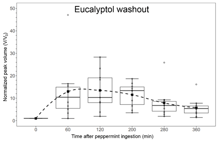
Figure 1. Eucalyptol washout measured via the micro-thermal desorber-gas chromatograph-ion mobility spectrometer (µTD-GC-IMS).


Human breath volatilome: a comparison of sampling and mass spectrometry methods for biomarker discovery in clinical research


Elodie Lamy1, Camille Roquencourt2, Nicolas Hunzinger1, Philippe Devillier1, 2, Emmanuelle Bardin1, 2, 3, Julie Mercier1, Stanislas Grassin-Delyle1, 2


1Université Paris-Saclay, UVSQ, INSERM, Infection et inflammation (2I), U1173, Département de Biotechnologie de la Santé, Montigny le Bretonneux, France

2Hôpital Foch, Exhalomics®, Suresnes, France

3Institut Necker-Enfants Malades, Paris, France


Background

Precision medicine lacks non-invasive biomarkers that can be measured using high-performance, rapid techniques. Volatilomics, an 'omics' approach, focuses on analyzing breath volatile organic compounds (VOCs). Mass spectrometry (MS) serves as the reference technology, but various types of mass spectrometers can be employed, leading to the generation of signals with diverse characteristics and levels of exhaustiveness. The aim of this study was to compare the volatilomic information obtained from online or offline mass spectrometry techniques in clinical studies.


Methods

We conducted a single-center clinical study in a university hospital (VOC-Compare, NCT06020521). Breath samples from 40 healthy participants were analyzed with three real-time MS instruments: PTR-Qi-TOF and PTR-TOF 10k (proton transfer reaction – mass spectrometry, Ionicon), as well as with a Soft Ionization by Chemical Reaction in Transfer (SICRIT®, Plasmion) ion source coupled to a Q-exactive MS (Thermofisher) (3 exhalations each). Breath was also collected on sorbent tubes (Tenax TA, Markes) using both a ReCIVA device (Owlstone) and sampling bags (Tedlar® bag, SKC) (0.5 L/tube) for subsequent thermal desorption and bidimensional gas chromatography – mass spectrometry (TD-GCxGC-MS) analysis (BT4D, Leco).


Results

A total of 120 breath samples were obtained. Using real-time analysis, 104 features were detected with the PTR-Qi-TOF (mass range 28-371 m/z), 252 with the PTR-TOF 10k (28-373 m/z), and 586 with the SICRIT-HRMS (51-294 m/z) after data filtering. High reproducibility was achieved reaching 7%, 6%, and 19% intra-participant variability, respectively.


GCxGC-MS provided 999 and 1144 filtered features (40-596 m/z) in samples collected with Tedlar® bags and ReCIVA, respectively. Annotated features accounted for 86% and 89% of detected features, respectively, with 34% features in common.


Conclusions

Each MS technique comes with its own set of advantages and limitations in terms of implementation and performance. The nature and completeness of the information obtained varies among them, suggesting potential benefits in selecting a technique based on its unique advantages or in combining several of them to enhance the completeness of the information generated during clinical studies aimed at discovering biomarkers.


Fundings

This work was supported by Région Île de France (VolatolHom, SESAME 2016 and MeLoMane, DIM 1HEALTH 2019), Saint-Quentin en Yvelines (ESR 2021) and Fondation Foch (VolatolHom and VOC-Info).


A state-of-the-art laser technology for detecting breath molecules


Qizhong Liang and Jun Ye


JILA


A laser technology built upon Nobel-winning technology (2005, Physics) has recently been tested its utility for breath-based diagnostics for the first time [Liang et al 2023 JBR; Liang et al 2021 PNAS]. In a trial study over 170 research subjects, breath tests using this laser technology reported a matching rate of 85% to the standard RT-PCR tests. In this talk, we will give a general introduction to its working principle, showing how we can detect multiple molecular species in a breath sample free from chemical reactions and down to parts-per-trillion detection sensitivity.


Defining VOC Signatures of Airway Epithelial Cells After Environmental Exposures


Angela M. Linderholm1,2, Kat Aribindi1, Eva Borras3, Katie Hamera3, Keith Bein5, Mitchell M. McCartney3,4, Cristina Davis3,4, Richart W. Harper1,2,4, Nicholas J. Kenyon1,2,4


1Division of Pulmonary, Critical Care and Sleep Medicine, University of California, Davis, Sacramento, CA

2Lung Center, University of California, Davis, Davis, CA

3Mechanical and Aerospace Engineering, University of California, Davis, Davis, CA

4VA Northern California Health Care System, Mather, CA

5Air Quality Research Center, University of California, Davis, Davis, CA


Background

We proposed to identify volatile organic compounds (VOCs) produced by the lung upon exposure to environmental pollutants to develop an exhaled breath signature that will be utilized for future clinical studies. Previously, we developed a reliable method to measure VOCs emitted from well-differentiated tracheobronchial epithelial cells in vitro. Using this method, we exposed well-differentiated small airway and bronchial airway epithelial cells to varying doses of traffic-related air pollutants (TRAP) to determine specific VOC signatures after exposure to TRAP. We utilized TRAP collected from the Caldecott tunnel in Oakland, CA to model “real-life” exposures. 


Methods

Human bronchial/tracheal airway epithelial (BAE) cells and small airway epithelial (SAE) cells were obtained from Lifeline Cell Technology (Frederick, MD).  The BAE and SAE cells were plated on Transwell (Corning Costar, Corning, NY) chambers (12 mm) at 1–2 × 10⁴ cells/cm2 coated with 0.05mg/mL type IV collagen (Sigma) in the PneumaCult-Ex medium (Stemcell Technologies). Once BAE and SAE cultures were confluent, they were transferred to ALI culture conditions in their respective media (Stemcell Technologies) for 1 month. Transwells containing confluent cells were placed into glass jars filled with 5mL of the appropriate media and capped with lids that had Twisters magnetized to them. The VOCs were extracted from the Twisters and analyzed using mass spectrometry. We also collected media samples to measure IL-6 and IL-8 protein levels secretion by ELISA.


Results

Exposure to TRAP resulted in distinct VOC and IL6 and IL8 responses, that differed between BAE and SAE cells, as well as individual subjects with little overlap. The VOCs produced by cells included aldehydes, ketones and hydrocarbons that previously have been linked to oxidative stress pathways.


Conclusions

Our studies suggest that TRAP exposure induced a specific cellular response, unique to BAE and SAE cells, related to their different functions that can be exploited for future clinical studies. We will further explore this phenomenon in future in vitro studies and pursue identification of the VOCs to assess metabolic pathways of importance.


Chip-Scale Mass Spectrometry for Point-of-Care Breath Diagnostics


Spiros Manolakos, Ashish Chaudhary, Ph.D., Kelli Barr, Ph.D., Christina Davis, Ph.D.


Detect-ION, University of South Florida, University of California - Davis


Background

The recent COVID-19 pandemic has underscored the urgency of swiftly identifying infected individuals to intervene early and prevent the progression to severe illness, as well as to curb the transmission of infections within communities. Exhaled breath diagnostics can provide such rapid screening approach while being non-invasive, cost-efficient, and potentially adaptable to detect various infectious agents, sometimes even before symptoms appear. Traditional breath analysis methods often entail collecting breath samples using tubes or bags for transportation to labs where Gas Chromatography-Mass Spectrometry (GC-MS) is employed for detecting trace-level organic compounds. While considered the gold standard, this method is both costly and logistically cumbersome. To address this, Detect-ION has leveraged its cutting-edge "chip scale mass spectrometry" technology, enabling a compact 10-L Preconcentrator-Thermal Desorption-Gas Chromatograph-Mass Spectrometer (TD-GC-MS) system, called “CLARION”, for analyzing exhaled breath.


Objective

In CLARION, our goal is to identify the relevant VOC biomarkers in exhaled breath that distinguish between infected individuals and a healthy population. If successful, a single CLARION device could require no more than a 1-minute breath sample per individual and conduct up to 160 breath analyses per day. Moreover, this approach could be more cost-effective than rapid PCR tests, enabling early detection of infections and facilitating high-throughput screening in large populations.


Methods

In CLARION project, we have established an institution review board (IRB) and plan to enroll asymptomatic and symptomatic human subjects across two sequential breath collection campaigns of cohort sizes of 100 (Campaign-1) and 500 (Campaign-2) respectively. Human subjects enrolled for the study will provide breath specimens, as well as nasal and throat swabs. Rapid antigen tests will evaluate the specimens for influenza, SARS-CoV-2, RSV, and group A streptococcus. In addition, RT-PCR will also be performed as a secondary diagnostic to validate antigen testing, with plans to perform BioFire® Respiratory 2.1 panel to accurately detect and identify the pathogens most associated with respiratory infections. Breath samples will be collected into the portable CLARION device for analysis, and into Tedlar bags and sorbent tubes for laboratory analysis on a commercial benchtop GC-MS. GC-MS data from the CLARION platform will be processed to generate a fingerprint pattern consisting of calibrated retention times and chemical identities for each VOC peak. These patterns along with the controls, which categorize each subject as uninfected or infected, will be provided as input to a Partial Least-Squares Discriminant Analysis (PLSDA) model building tool to develop detection algorithms.


Preliminary Results

The IRB has been accepted and we plan to begin enrollment for Campaign-1 in June 2024. A miniature breath collector with an embedded thermal desorption stage has been developed and

successfully integrated into the CLARION platform. The MBC has smart collection technology to perform volumetric sampling of breath. Initial studies of vapor phase analytical standards have shown sensitivity of the system in the low parts-per-billion to high parts-per-trillion range.


Conclusion

Fieldable GC-MS system adapted to collect exhaled human breath can provide a versatile diagnostics capability for rapid screening of a large population.

From research to routine: Meeting the evolving demands of clinical breath analysis with advancements in thermal desorption


Helen Martin1, Laura Miles1, Ryan Francis1, James Swift2 and Matthew Turner2


1Markes International Ltd, 1000B Central Park, Western Avenue, Bridgend, CF31 3RT, UK

2Department of Chemistry, Loughborough University, Loughborough, LE11 3TU, UK


Breath-based biomarker discovery research is entering a particularly exciting phase with many studies scaling up to large clinical trials and generating viable candidate marker compounds. As the size of sample sets increases, so too does the duration of individual studies and the number of personnel involved. This scale-up of efforts introduces more opportunities for error and highlights the need for rigorous quality control.


Thermal desorption (TD) coupled with gas-chromatography mass-spectrometry (GC-MS) has long been established as the gold standard technique for breath analysis. Volatile organic compounds (VOCs) in breath are pre-concentrated by collection onto sorbent packed tubes and shipped to a central laboratory for analysis. These tubes are small, physically robust, easy to transport and provide extended sample storage stability compared to other methodologies.


In this presentation we will discuss how quality control strategies can be built into thermal desorption-based workflows throughout the life cycle of every sample tube. These strategies include the use of leak testing, surrogates and internal standards and automated water management. We will explore the benefits of re-collecting a portion of breath samples for further analyses and show how this re-collection can reduce the sampling burden, aid sample archiving and extend the dynamic range of the analysis to help manage the wide-ranging concentration of important breath components.


We will also review the generation of pooled biological quality control samples using re-collection and how they could be applied to proficiency testing and longitudinal studies. Pooled quality control samples are a core concept in metabolomic studies, providing a representative sample matrix and metabolite composition. However, these very useful and informative samples are not easily produced or integrated into breath VOC workflows. To demonstrate the concept of a pooled quality control sample for breath, we utilise the protocol described in the peppermint experiment (Henderson et al 2020). Here, multiple breath samples are collected over a 10-hour period after the ingestion of a peppermint oil capsule. The resulting breath samples contain several VOCs including menthol, menthone and alpha-pinene which exhibit a range of concentrations in different participants at the various time points.


We will explore how these pooled breath samples can be applied to routinely evaluate data quality, reproducibility of the method and instrumentation, and correct for any system bias over time.


1.     Ben Henderson et al 2020 J. Breath Res. 14 046008


Selectivity to diagnose COVID-19 infection from exhaled breath volatiles (EBV) against other common upper respiratory infections


Mitchell M McCartney, Eva Borras, Dante E Rojas, Tiffany Lam, Enrique Lopez, Nicholas J Kenyon, Cristina E Davis


Mechanical and Aerospace Engineering; UC Davis Lung Center; and the Department of Internal Medicine, UC Davis, Davis, California. VA Northern California Healthcare System, Mather, California.


Background

Many studies have reported that COVID-19 infection can be identified through metabolites in exhaled breath vapor (EBV). However, during the first two years of the COVID pandemic, there was a stark decline in incidences of other respiratory infections, such as influenza and RSV, which nearly disappeared globally. Without transmission of non-COVID pathogens, it was not possible to compare the selectivity of a breath-based test to identify COVID+ patients against other common upper respiratory infections. Thus, it was unknown if the reported shift in EBV from COVID-19 was a unique signature to COVID, or whether COVID breath tests might be confounded by a general response to airway infection as observed in breath.


Objective/Aims

The objective of this study was to collect EBV samples from patients with known respiratory infections, including coronaviruses, influenzae, and RSV, to determine the selectivity of a breath-based test to diagnose COVID-19.


Methods

Breath vapor samples were collected from patients exhibiting symptoms of an upper respiratory infection. A nasal swab was also collected and analyzed using the BioFire Respiratory 2.1 Panel, which targets 22 viruses and bacteria, including SARS-CoV-2. Those positive for SARS-CoV-2 were phenotyped, as we previously reported that major shifts in COVID variant (eg, from Delta to Omicron) significantly altered the COVID breath signature.


Collection occurred at the UC Davis Medical Center and from the surrounding Sacramento community under an IRB-approved protocol, #1636182. Demographic information about volunteers was collected through a questionnaire and review of electronic medical records.


Breath was collected in Tedlar bags, then extracted onto Tenax TA sorbent tubes. A paired background/environmental air sample was collected for each breath sample to remove exogenous features from the corresponding breath sample.


Samples underwent thermal desorption-gas chromatography-mass spectrometry (TD-GC-MS) analysis. GC-MS data were deconvoluted and aligned. Background VOCs were subtracted from breath samples. The dataset was randomly split into a training and validation set for machine learning algorithms to identify the COVID-19 breath signature.


Results / Conclusion

At time of abstract submission, sample collection and chemical analysis are ongoing, with 177 negative controls enrolled, 147 COVID+, 4 influenza, 2 rhinovirus, and 47 samples from symptomatic patients pending analysis of their nasal swab. During the IABR Meeting, the authors will present the latest findings, detailing the accuracy, sensitivity and specificity of a breath-based GC-MS assay to diagnose COVID-19 against other respiratory pathogens. We will present findings on whether COVID-19 biomarkers correlated with demographic factors such as age, ethnicity, or correlated with disease severeness and symptoms.


Peptides and nanotubes - a machine learning and biological approach to VOC sensing


Oliver Nakano-Baker, Richard V. Lee, Shalabh Shukla, J. Devin MacKenzie


University of Washington Materials Science & Engineering, University of Washington Laboratory Medicine & Pathology, University of Washington Mechanical Engineering


Background

Volatile organic compounds (VOCs) in breath are promising clinical biomarkers for various diseases, but detecting VOCs outside of the lab presents challenges. Portable eNoses struggle to match the sensitivity and specificity of mass spectroscopy to key VOC markers of disease. We recently demonstrated an alternate approach to portable breath fingerprinting: a bioelectronic platform that combines carbon nanotube (CNT) sensors with surface-binding peptide molecules derived from olfactory proteins.

Objective: Here, we propose a pipeline to design, combine, and fabricate micro-scale biomimetic sensors for specific VOCs. These sensors are the building blocks of eNoses that are capable of performing targeted breath-based diagnostics. Ideally, the pipeline translates a VOC fingerprint of a disease to a fabricated multiplex chip capable of detecting the fingerprint. It is demonstrated here by creating a sensor for ethyl butyrate, a VOC marker of bacterial infections and COVID-19 disease.

Methods: Sensor design proceeds in three steps. First, bioinformatics and molecular dynamics identify peptide probes with binding affinity to certain VOCs, creating a library of targeted binders. Second, bootstrapped machine learning models evolve the peptide composition of a multi-channel eNose to maximize the ability to classify disease fingerprints. Finally, CNT field-effect transistors are incubated with the engineered peptides to sense VOCs by direct signal transduction with sub-parts-per-billion sensitivity.


Results

A multiplex sensor consisting of just 5 peptide-CNT transistors is capable of identifying the VOC fingerprint of COVID-19 in the bootstrapped design loop, and a 10-plex design extends this capability to multiple respiratory diseases, including influenza, rhinovirus, and asthma. All engineered probes display selective binding to their VOC targets as measured by quartz crystal microbalance, and a CNT transistor utilizing a peptide probe that targets the VOC ethyl butyrate exhibits p&lt;0.001 stronger source-drain current when its target is present, compared to a control at the same concentration.


Conclusion

With promising analytical performance and an expansive probe design space, the biomimetic eNose promises a step-change in the sensitivity and specificity of micro-scale VOC detection. If challenges of durability and human variability can be addressed, it could be the key to mobile breathomics - phone-size breath sensors capable of multi-viral diagnoses, blood glucose monitoring, cancer screening, and more.


Disclosures

Research was performed at the University of Washington (UW) under the WE-REACH program through the RADx RAD program at NIDCR/NIH and the Washington Research Foundation. Additional funds were provided by UW CoMotion, the School of Dentistry, and NSF grant 2313269. Authors Nakano-Baker, Lee, and Shukla are co-founders of Odo Labs, LLC.


From dynamic nature to human origin of breath isoprene – an investigative story


Pritam Sukul1, Anna Richter2, Christian Junghanss2, Jochen K Schubert1, Wolfram Miekisch1


1Rostock Medical Breath Research Analytics and Technologies (ROMBAT), Dept. of Anesthesiology, Intensive Care Medicine and Pain Therapy, University Medicine Rostock, Rostock, Germany,

2Department of Medicine, Clinic III – Hematology, Oncology, Palliative Medicine, Rostock University Medical Center, Rostock, Germany


Background

Isoprene (1,3-pentadiene, C5H8) is amongst the most abundant volatile metabolites produced by plants and animals. In humans, endogenously produced isoprene is exhaled in nearly everybody’s breath in high amounts and has been described as a potential biomarker for various diseases1. Exhaled isoprene is extremely dynamic in nature2 and concentration changes are also related to physio-metabolic, inherited conditions and ageing3–5. Based on pre-clinical in vitro experiments, Deneris et al hypothesized that cholesterol biosynthesis in the liver potentially gives rise to isoprene in our breath6. Nevertheless, changes and/or differences observed in breath isoprene under various conditions investigated in clinical studies could not be justified by the suggested origin. Thus, missing knowledge of the origin and pathways involved hindered the translation of this biomarker into clinical practice. Isoprene absent healthy adults is rare (<0.3/%) in the nature. Our previous investigations in such a healthy German adult and her family (isoprene deficient) disqualified the putative origin and depicted a recessive inheritance of this phenotype7.


Objective

We aimed to find the human metabolic origin of isoprene8.


Methods

In consecutive clinical breath screening studies, amongst 2000 subjects, we identified five healthy German adults without breath isoprene. After having the ethical clearance, we conducted venous blood collection, peripheral blood mononuclear cells isolation, DNA isolation, multi-omics (breathomics, genomics) and serological metabolites analysis in healthy isoprene aberrated and isoprene normal adults8.


Results

We obtained five (3 females and 2 males) isoprene absent healthy adults, 64 (39 females and 25 males) isoprene deficient (with low exhaled concentrations <50ppbV). Whole exome sequencing in these individuals revealed only one shared homozygous (<1% prevalent in Europeans) IDI2 stopgain mutation (at c.431 position) causing loss of enzyme active site and Mg–cofactor binding sites and thereby, preventing conversion of isopentenyl diphosphate to dimethylallyl diphosphate (DMAPP) in cholesterol metabolism. Targeted sequencing depicted that the IDI2 variant is heterozygous in isoprene deficient blood-relatives and absent in unrelated isoprene normal adults.  Wildtype IDI1 and cholesterol metabolism related serological parameters were normal in everyone. Unlike other mammals, naturally IDI2 knocked out pigs and bottlenose dolphins exhale no isoprene. Human IDI1 is expressed highly in liver but the hepatocellular cytochrome P450 enzymes immediately oxidizes isoprene. Skeletal muscles metabolize lipids (oxidize cholesterol, fatty acids) to produce energy, regulate intramyocellular signalling and integrity. Peroxisomal beta-oxidation produces acetyl-CoA i.e., channelled towards DMAPP production. Thus, human IDI2 determines isoprene production as DMAPP is the only source of isoprene and unlike plants, humans lack isoprene synthase and its enzyme homologue. Human IDI2 is only expressed in skeletal-myocyte peroxisomes and instant spikes in isoprene exhalation during any muscle activity underpin the origin from muscular lipolytic cholesterol metabolism.


Conclusions

Our discovery of the genetic origin and metabolic routes of human isoprene production enabled objective interpretations and applications of isoprene as a noninvasive biomarker. Knowledge of accurate metabolic source and normal/dynamic range are indispensable for valid clinical interpretation of any endogenous marker. Besides isoprene, our breath contains many potentially endogenous VOCs and their origins are yet uncertain.
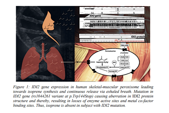
Graphic from the abstract, "From dynamic nature to human origin of breath isoprene – an investigative story "

References

1. Mochalski, P., King, J., Mayhew, C. A. & Unterkofler, K. A review on isoprene in human breath. J. Breath Res. (2023) doi:10.1088/1752-7163/acc964.

2. Miekisch, W., Sukul, P. & Schubert, J. K. Chapter 2 Origin and Emission of Volatile Biomarkers in Breath: Basics and Dynamic Aspects. in Volatile Biomarkers for Human Health: From Nature to Artificial Senses 22–38 (The Royal Society of Chemistry, 2023). doi:10.1039/9781839166990-00022.

3. Sukul, P. & Trefz, P. Physio-Metabolic Monitoring via Breath Employing Real-Time Mass Spectrometry: Importance, Challenges, Potentials, and Pitfalls. in vol. 4 1–18 (Springer, 2022).

4. Hoffmann, G. et al. Mevalonic Aciduria — An Inborn Error of Cholesterol and Nonsterol Isoprene Biosynthesis. New England Journal of Medicine 314, 1610–1614 (1986).

5. Sukul, P. et al. Physiological and metabolic effects of healthy female aging on exhaled breath biomarkers. iScience 25, 103739 (2022).

6. Deneris, E. S., Stein, R. A. & Mead, J. F. Invitro biosynthesis of isoprene from mevalonate utilizing a rat liver cytosolic fraction. Biochemical and Biophysical Research Communications 123, 691–696 (1984).

7. Sukul, P., Richter, A., Schubert, J. K. & Miekisch, W. Deficiency and absence of endogenous isoprene in adults, disqualified its putative origin. Heliyon 7, e05922 (2021).

8. Sukul, P., Richter, A., Junghanss, C., Schubert, J. K. & Miekisch, W. Origin of breath isoprene in humans is revealed via multi-omic investigations. Commun Biol 6, 1–12 (2023).


Developing novel perovskite-based nanomaterials for the detection of volatile biomarkers in exhaled breath


Y.L. Pham1, J. Beauchamp1, A. Kostopoulou2, K. Brintakis2, E. Stratakis2, L. Manna3


1Fraunhofer Institute for Process Engineering and Packaging IVV, Giggenhauser Str. 35, 85354 Freising, Germany.

2Foundation for Research and Technology – Hellas (FORTH), Ν. Plastira 100, Vassilika Vouton, GR - 700 13, Heraklion, Greece.

3Fondazione Istituto Italiano di Tecnologia (IIT), Via Morego, 30, 16163 Genoa, Italy.


Abstract

Comprehensive laboratory-based analytical devices pose limitations due to the skills and training required for operation, rendering it impractical for routine use in breath testing. Additionally, these devices are not suitable for therapeutic monitoring or wearable diagnostic applications. Therefore, there is a pressing need to miniaturize breath analysis sensors to achieve a low-cost solution with high sensitivity, selectivity, and rapid response time for detecting volatile organic compounds (VOCs) at low concentrations. However, the development of breath-sensing materials is a complex task that demands expertise in both materials development and breath analysis. This duality is essential for creating proof-of-concept sensors for detecting exhaled breath markers for targeted applications.


A current interdisciplinary European study aims to promote new opportunities for participating partners (FORTH, IIT, FHG) to improve the levels of excellence and expertise of all three institutions in the fields of novel perovskite nanocrystals for sensing applications and breath-based diagnostics. Over the course of the project, perovskite sensing elements are synthesized and characterized, which are imperative for the fabrication of breath sensing devices. Currently, different structures of perovskite-based nanocrystals are tested for the detection of selected VOCs. The sensing ability for breath VOCs will be then evaluated in simulated conditions using different target volatiles relevant to the peppermint experiment1, 2 – a benchmarking initiative established by the International Association of Breath Research (IABR) that seeks to address the lack of inter-comparability of outcomes across independent breath biomarker studies. Once established, this system will be implemented in the peppermint experiment with real breath samples to obtain benchmark values for the perovskite-based nanomaterials to establish this technology in breath research.


The poster outlines the project concept for the development and validation of novel breath-sensing devices, the materials and methods being employed, and intends to spark debate on novel perovskite-based nanomaterials for the detection of breath volatiles.


References

1Henderson, B. et al. A benchmarking protocol for breath analysis: the peppermint experiment. J. Breath Res.14.4 (2020): 046008.


2Pham, Y. L., Yu, R., Beauchamp, J. Cross-validation of the peppermint benchmarking experiment across three analytical platforms. J. Breath Res. 17 (2023): 046003.


Interest of Soft Ionization by Chemical Reaction in Transfer (SICRIT) – high resolution mass spectrometry for exhaled breath analysis in clinical studies


Camille Roquencourt1, Elodie Lamy2, Nicolas Hunzinger2, Hélène Salvator3, 4, Philippe Devillier1, 4, Emmanuelle Bardin1, 2, 5, Stanislas Grassin-Delyle1, 2


1Hôpital Foch, Exhalomics®, Suresnes, France

2Université Paris-Saclay, UVSQ, INSERM, Infection et inflammation (2I), U1173, Département de Biotechnologie de la Santé, Montigny le Bretonneux, France

3Service de pneumologie, Hôpital Foch, Suresnes

4Laboratoire de recherche en Pharmacologie Respiratoire – VIM Suresnes, UMR 0892, Université Paris-Saclay, Suresnes, France

5Institut Necker-Enfants Malades, Paris, France


Background

Real-time mass spectrometry (MS) breath analysis is a non-invasive, rapid method for disease diagnostic and treatment monitoring that relies on the detection of volatile organic compounds (VOC). A novel approach for real-time VOC analysis involves an ambient flow-through ionization technique, using Soft Ionization by Chemical Reaction in Transfer (SICRIT) - mass spectrometry with atmospheric pressure inlet.


Objective

The objective of the study was to assess the feasibility of implementing exhaled breath analysis by SICRIT-MS within the context of clinical studies.


Methods

We conducted a clinical study within a university hospital (VOC-Compare, NCT06020521). Main inclusion criteria were adult healthy volunteers and non-inclusion criteria were pregnancy, any chronic condition and smoking. Measurements included breath analysis with SICRIT-MS (Plasmion), among other techniques. SICRIT parameters were set at 1600 V and 15000 Hz. VOC detection was carried out using a Q-Exactive mass spectrometer (Thermofisher) in positive ionization mode, with full-scan data acquisition in the range 50-300 m/z. Sheath and auxiliary gas flow rates were set at 0 and 12 arbitrary units, respectively. The capillary temperature was maintained at 200°C, and the S-Lens RF level was set at 65. Participants performed three consecutive expirations through disposable mouthpieces into the device, aiming to sustain an expiration airflow at 8 L/min, which was simultaneously assessed in real-time. The primary outcome measure was the number of reliable individual signals detected. Data analysis was performed using an in-house R script, involving mass calibration, peak detection in the mass dimension at each time point, alignment of peaks in the time dimension, and the selection of compounds specifically detected in the expiration phases.


Results

Breath samples from 40 healthy participants were successfully analyzed. Preliminary data analysis was conducted, and expiration phases were accurately identified using isoprene as a tracer (m/z 69.07). Calculated resolution on the three calibration peaks (m/z 67.069, 149.044 223.063) was in the range of 75,000 to 130,000. The total average feature number per sample was 1090, with 59% of features significantly more abundant in expiration phases than in the background. After alignment and isotope suppression, a total of 586 features (mass range: m/z 51-294, mean signal to noise ratio: 45) were detected in more than 30% of participants' breath samples, including 417 features detected in the breath of all individuals.


Conclusion

Our results demonstrate the successful implementation of SICRIT-MS breath analysis in clinical studies, highlighting its potential as a means for real-time biomarker discovery in breath utilizing high-resolution mass spectrometry detection.

Investigating Exhaled Volatile Organic Compounds in Healthy Breath Using Solid Phase Microextraction and Gas Chromatography-Mass Spectrometry


Eray Schulz1,2, Shivaum Heranjal3, Mariana Maciel1, Sha Cao4, Xiaowen Liu5, Mark Woollam1,2, and Mangilal Agarwal1,2,3,6


1Chemistry and Chemical Biology, Indiana University Indianapolis, IN, United States.

2Integrated Nanosystems Development Institute, Indiana University Indianapolis, IN, United States.

3Electrical and Computer Engineering, Purdue University Indianapolis, IN, United States.

4Biostatistics and Health Data Science, Indiana University School of Medicine, Indianapolis, IN, United States.

5Biomedical Informatics and Genomics, Tulane University School of Medicine, New Orleans, LA, United States.

6Biomedical Engineering and BioHealth Informatics, Luddy School of Informatics, Indiana University Indianapolis, IN, United States.


Background

Volatile organic compounds (VOCs) in exhaled breath are potential biomarkers that may facilitate non-invasive health monitoring in diverse locations including at-home. One challenge that still exists however, is that baseline levels of VOCs in relatively healthy breath have not been benchmarked.


Objective

This study aimed to identify baseline levels of VOCs in exhaled breath using solid phase microextraction (SPME) and gas chromatography – mass spectrometry (GC-MS).


Methods

An optimized and standardized direct breath sampling method using solid-phase microextraction (termed DB-SPME) coupled to gas chromatography – mass spectrometry (GC-MS) was implemented to analyze samples collected from 164 “healthy” volunteers. 10 of these volunteers provided an additional 9 samples longitudinally over the course of 6 months. Blood samples for comprehensive metabolic panels and complete blood counts were collected, as well as data from a food diary and lifestyle questionnaire to obtain information on confounding variables that may impact “healthy” VOC levels.


Results

After instrumental analysis, data processing procedures were implemented (removing SPME artifacts, subtracting background air, etc.) which identified a total of 62 VOCs including terpene/-oids, carbonyls, aromatics and other functional groups. Hierarchical clustering showed cyclic monoterpenes had similar trends in healthy breath samples with a significant correlation of specific monoterpenes including α-pinene and β-pinene (R2=0.58, p=1x10-29), pointing to a relationship between structure and expression. Reproducibility was also benchmarked, showing cross-sectional VOC measurements were significantly more variable compared to those collected longitudinally. Correlation analyses of VOCs and confounding variables showed male volunteers had significantly elevated levels of breath isoprene relative to women (p=0.04), corresponding to previous literature. Along with this, acetone and isoprene breath signals were quantified using calibration curves developed from a previous study. Acetone and isoprene concentrations ranged from 21ppb to 773 ppb and 58ppb to 1734ppb in the cross-sectional cohort respectively. Longitudinal concentration ranges were lower, with acetone being between 22ppb-343ppb, and isoprene between 38ppb-983ppb.


Conclusion

In the future, the results from this work can be leveraged to solidify our understanding of baseline levels of VOCs in breath that can be applied to disease-specific studies. Furthermore, continuing to quantify VOC signals can help push the field of breath biopsy to more clinically translatable applications.


Conflict of Interest

Mangilal Agarwal has an ongoing collaboration with the NANOZ company and Scosche Industries to commercialize sensors to detect VOCs presented in this work for noninvasive health monitoring applications. All other authors report no conflicts of interest relevant to this article.


Ethics Board Approval

All subjects provided written consent to participate in this study, and Institutional Review Board (IRB)/Ethics Committee approval was obtained (IRB # 12954). Institutional Biosafety Committee (protocol #IN-1301) approval was also obtained at Indiana University (IU).


Extending Laser Absorption Spectroscopy towards Detection of Larger Volatile Organic Compounds in Breath


Miloš Selaković1,2, Lukas Emmenegger1, Renato Zenobi2, Béla Tuzsona1


1Laboratory for Air Pollution/Environmental Technology, Empa, Überlandstrasse 129, 8600 Dübendorf, Switzerland

2Department of Chemistry and Applied Biosciences, ETH Zürich, Vladimir-Prelog-Weg 1−5/10, 8093 Zürich, Switzerland


Background

Laser absorption spectroscopy (LAS) is well-established for trace gas measurement of small, inorganic molecules, providing fast and accurate response in relatively compact, easy-to-use, and cost-effective instrumentation. Extending LAS towards the detection of volatile organic compounds (VOCs) would be highly beneficial for future point-of-care diagnostics based on human breath. However, larger VOCs often exhibit broad spectral absorption features which are usually attributed to the overlap of a multitude of absorption lines from many vibrational modes, and therefore limiting the LAS to detection of VOCs with molar mass <40 g/mol. [3] Despite this common view, we recently developed a compact mid-infrared (mid-IR) analyzer for simultaneous measurement of small VOCs in breath. [1,2] In this work, we show that even larger molecules (with a molar mass over 100 g/mol) can be measured by LAS.


Methods

We used a custom-built spectrometer based on a widely electrically tunable quantum cascade laser that allows access to six different spectral windows around 1080 cm–1 wavenumber. [1] VOC gas standards were generated in the laboratory by vaporization of liquid samples into a carrier gas stream in a dynamic injection design [4]. The samples were analyzed in a flowthrough configuration at low gas pressure (~50 mbar) to reduce the broadening of the absorption lines. The high-resolution spectra of pure substances were used for the generation of a reference database.


Results

By extensive validation of our method through the series of spiking experiments, we demonstrated that our instrument is well-suited for the quantification of oxygen-containing VOCs at amount fractions down to tens of ppb. The broad measuring range, high spectral resolution, and the unique spectral fingerprints of the investigated VOCs assure excellent selectivity of the method and enable multi-compound measurements in the breath. With a time resolution of 360 ms, concentration profiles of several VOCs, water and CO2 in one breath stroke can be retrieved with a relative expanded uncertainty (k=2) of <2%.


Spectral screening of over 50 different VOCs revealed significant fine structure not only in the ro-vibrational spectrum of small (up to 4 non-hydrogen atoms) but also of large molecules (6 or more non-hydrogen atoms) with a rigid and symmetrical chemical structure. To investigate the effect of molecular symmetry and rigidity on spectral fine structure in mid-IR, several exemplary compounds with similar molar mass but different structures have been investigated. One such example is shown in Figure 1. We argue that the absence of spectral fine structure in heavy linear-chain low-symmetry molecules is related to a large number of available con- formers where each conformer contributes to the complexity of the spectrum. We demonstrated that our method can capture different conformers of a compound in the example of Nmethylformamide. These findings have the potential to substantially increase the number of possible VOCs that can be analyzed with LAS and open up the path towards other applications, such as conformational analysis.

Graphs from Breath Summit 2024 abstract, "Extending Laser Absorption Spectroscopy towards Detection of Larger Volatile Organic Compounds in Breath" depicting absorption spectra of butyric acid ( on left) and 1,4-dioxane (on right). 1,4-dioane has a constrained ring structure and shows distinct features that are well-resolved at reduced gas pressure (right).

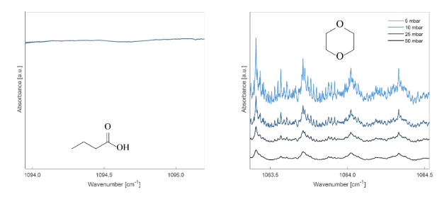
Figure 1. Absorption spectra of butyric acid (left) and 1,4-dioxane (right). 1,4-dioane has a constrained ring structure and shows distinct features that are well-resolved at reduced gas pressure (right).


Conclusion

We demonstrated that LAS can be applied for the analysis of some larger VOCs. Our findings may initiate a paradigm shift in the analysis of organic molecules by mid-IR laser spectroscopy, in particular for breath analysis.


Acknowledgements

We acknowledge support from Zürich Exhalomics [5] and Evi Diethelm-Winteler-Stiftung.


References

[1] R. Brechbühler et al. Anal. Chem. 2023, 95, 2857

[2] M. Selaković et al. Chimia 2023, 77, 785

[3] J. Wojtas et al. Opto-Electronics Rev. 2012, 20, 26.

[4] W. Vautz and M. Schmäh Int. J. Ion Mobil. Spec. 2009, 12, 139

[5] https://www.exhalomics.ch/ (Accessed: 15.01.2024)

Diagnosing Pulmonary Tuberculosis in Children through Breath Analysis: An Emerging Method


Ning Sun1, Carly A. Bobak3, Lesley Workman4, Lindy Bateman4, Margaretha Prins4, Cynthia Baard4, Mark P. Nicol5, Heather J. Zar4 & Jane E. Hill1,2*


1School of Biomedical Engineering, University of British Columbia, Vancouver, BC, Canada

2Department of Chemical and Biological Engineering, University of British Columbia, Vancouver, BC, Canada

3Department of Biomedical Data Science, Dartmouth College, Hanover, New Hampshire, USA

4Department of Pediatrics and Child Health, MRC Unit on Child and Adolescent Health, University of Cape Town and Red Cross War Memorial Children’s Hospital, Cape Town, South Africa

5School of Biomedical Sciences, University of Western Australia, Perth, Australia


Background

Tuberculosis (TB) poses a significant threat to children, causing an estimated one million cases and 250,000 deaths annually. Diagnosis in children may be challenging due to nonspecific clinical symptoms, inability to spontaneously produce sputum, and low bacillary burden. Only about 30% of childhood TB cases are notified and diagnosed, with approximately 50% not having bacterial confirmation. Volatile molecules (VCs) in breath reflect alterations in the body's health status resulting from the host and/or the bacterium, as well as the microbiome. Utilizing VCs found in the exhaled breath of adults with TB disease holds promise as a triage test so that it may offer a non-invasive alternative for diagnosing pulmonary TB (PTB) in children.


Aim

(A) To investigate whether a subset of molecules in the breath of children with PTB has diagnostic utility. (B) To track the breath VCs of pediatric subjects during six months of treatment.


Methods

Age- and gender-matched children were prospectively enrolled in a TB diagnostic study in Cape Town, South Africa, and breath samples were collected. The disease was categorized as confirmed PTB (microbiological diagnosis), unconfirmed (clinically diagnosed), and unlikely TB (non-TB respiratory tract illness). From April 2017 to December 2017, 31 children were enrolled in an initial cohort, with one breath sample collected before the commencement of treatment. From December 2017 to September 2023, a further 143 children were enrolled and followed. Breath samples were collected before treatment and at 1, 3, and 6 months followup. The studies followed all protocols approved by the Institutional Review Board.


Breath was collected from each child using a straw, directed into a 1L Tedlar bag, and then concentrated onto a thermal desorption (TD) tube. The TD tube was capped and stored at 4°C until analysis. Breath analysis was conducted via two-dimensional gas chromatograph tandem time of flight mass spectrometry (GC×GC-ToFMS). Chromatographic data were aligned, and volatile compounds were assigned putative names based on spectral library match using ChromaTOF software. Statistical analyses and putative biomarker discovery using machine learning tools were performed in R.


Key Results

(A) In the initial cohort (10 confirmed TB, 11 unconfirmed TB, and 10 unlikely TB with a mean age of 6.0 ± 3.1 years), a set of four breath biomarkers was evaluated in characterizing the three mentioned statuses, achieving a sensitivity of 80% and a specificity of 100% (Fig.1). (B) In the second cohort (59 confirmed TB, 57 unconfirmed TB, and 27 unlikely TB with a mean age of 7.3 ± 3.3 years), a list of 116 molecules that contributed to the alterations in breath profiles during treatment was tracked from a subset of subjects. (C) Among them, two out of four published breath biomarkers were identified, of which 4- methyl octane significantly increased after treatment. Other molecules were selected to reliably predict the treatment at 1, 3, and 6 months, respectively (Table 1).


Conclusions

A set of breath biomarkers can discriminate PTB from other lower respiratory illnesses in children. Breath biomarkers also show promise in monitoring the TB treatment response.

Graphs from Breath Summit 2024 abstract, Diagnosing Pulmonary Tuberculosis in Children through Breath Analysis: An Emerging Method showing A set of breath biomarkers can discriminate PTB from other lower respiratory illnesses in children. Breath biomarkers also show promise in monitoring the TB treatment response.
 

Canine-Inspired Identification of Volatile Organic Compound Biomarkers for Noninvasive Health Monitoring: Opportunities in the 21st Century for Biotechnology


Mark Woollam1,2* and Mangilal Agarwal1,2,3


1Integrated Nanosystems Development Institute, Indiana University Indianapolis, IN, United States.

2Chemistry and Chemical Biology, Indiana University Indianapolis, IN, United States.

3Biomedical Engineering and BioHealth Informatics, Luddy School of Informatics, Indiana University Indianapolis, IN, United States.


*Presenting author; mwoollam@iu.edu


Background

Canines have the remarkable capability of smelling volatile organic compounds (VOCs) emanating from exhaled breath and other noninvasive biofluids to distinguish diverse medical conditions. Researchers around the world for decades have been exploring the chemical composition of breath and how this information can be utilized for biomedical applications. Patients with diseases including but not limited to diabetes, COVID-19, and different types of cancer can benefit from a noninvasive means of screening and diagnosis, and VOC profiling may be a promising avenue for exploration.


Objective

This research seeks to develop analytical methods to profile VOCs in exhaled breath with high sensitivity, and to use these techniques to identify specific analytes and biomarker panels for different medical conditions including COVID-19 and hypoglycemia. Strides are also taken to develop a more fundamental understanding of the VOC profiles in healthy breath along with their cross-sectional and longitudinal reproducibility.


Methods

Approximately 3L of exhaled breath was collected through a viral filter into a Tedlar bag to identify VOC biomarker panels for COVID-19 and hypoglycemia. VOCs collected in bags were transported to the laboratory, cryothermally transferred to headspace vials loaded with glass wool, and stored at -80℃ until analysis by solid phase microextraction (SPME) coupled to gas chromatography-mass spectrometry (GC-MS). For profiling VOCs in healthy breath, a direct breath sampling method (termed DB-SPME) was developed, standardized through capnography, and optimized for sensitivity. VOCs collected on to SPME fibers for both methods were analyzed using a GC-MS quadrupole time-of-flight system. Biostatistical analysis was undertaken on the collected data to identify biomarkers through univariate and multivariate approaches.


Results

VOC results for COVID-19 revealed that subjects with a positive diagnosis had a significantly higher number of compounds and total integrated signal. Individual analytes had relatively high accuracy alone, and pattern recognition through machine learning identified a biosignature of three VOCs that could distinguish COVID-19 with over 95% accuracy. Most interestingly, samples collected from those who recovered (two months after initial diagnosis) showed that the identified breath-based biomarkers are restored to healthy baseline levels. Chemometric analyses were also employed for hypoglycemia biomarker discovery, and a separate unique panel of six VOCs was identified that could detect hypoglycemia with sensitivity = 94.8% and specificity = 95.0%. When this model was blindly tested on independently collected samples, hypoglycemia was classified with sensitivity = 90.0% and specificity = 89.9%. Analyses of breath samples from a cohort of healthy volunteers were also accomplished, which showed cross-sectional VOC measurements were significantly more variable compared to those collected longitudinally. Additionally, correlation analyses of VOCs and confounding variables showed male volunteers had significantly elevated levels of isoprene relative to women (p=0.04).


Conclusion

The presented results can ultimately inspire the design and development of an integrated nanosensor array in the form of smart and connected breathalyzers that can be used for noninvasive health monitoring rapidly at a point-of-care.


Conflict of Interest

Mangilal Agarwal has an ongoing collaboration with the NANOZ company and Scosche Industries to commercialize sensors to detect VOCs presented in this work for noninvasive health monitoring applications. All other authors report no conflicts of interest.


Ethics Board Approval

All subjects provided written consent to participate in this study, and Institutional Review Board (IRB)/Ethics Committee approval was obtained (IRB #’s 12954, 2008193976). Institutional Biosafety Committee (protocol #IN-1301) approval was also obtained at Indiana University (IU).


Assessment of Exhaled Breath VOC Reproducibility on Tenax®GR Thermal Desorption Tubes


Darakshan Zabin1, Trenton J. Davis1,2, Ivan V. Ivanov3, Heather D. Bean1,2


1School of Life Sciences, Arizona State University, Tempe, AZ

2Center for Fundamental and Applied Microbiomics, The Biodesign Institute, Tempe, AZ

3Department of Veterinary Physiology & Pharmacology, Texas A&M University, College Station, TX


Background

We are conducting breath volatile organic compound (VOC) studies in partnership with research teams across North America, utilizing sorbent-packed thermal desorption tubes (TDTs) for breath VOC sample concentration, storage, and transport. Most of our studies are focused on identifying the differences in breath VOC profiles between groups of subjects (e.g., biomarkers that differentiate diseased vs. healthy cohorts), but within each cohort there are several sources of breath VOC variation that impact the study, including variation between subjects in a cohort, between breath samples within subject, and between TDTs. Additionally, storage time may also interact with these sources of variation and affect breath VOC sample reproducibility.


Objectives/Aims

In this study we aimed to quantify the reproducibility of breath VOC samples on TDTs within a subject and between subjects and assess the interaction of storage time with these sources of variation.


Methods

All procedures were approved by the U.S. Army Medical Research and Development Command (USAMRDC), Office of Human and Animal Research Oversight (OHARO), and Office of Human Research Oversight (OHRO; log numbers E03915.1a and E03915.1a-1). Four subjects were recruited to provide three breath samples in 10 L Tedlar bags. All breath samples were collected on the same day, and within 120 minutes of sample collection, each sample was split into 0.6 L technical replicates by transferring onto Tenax® GR TDTs. TDTs were stored at 4 °C and technical duplicate TDTs were analyzed after 0.5 to 32 days of storage using thermal desorption – comprehensive two-dimensional gas chromatography – time of flight mass spectrometry (TD–GC´GC–TOFMS). After data processing and alignment of 152 TDT breath samples, sample reproducibility and dissimilarity were assessed using pairwise distance metrics.


Results

On average, we found that within-subject breath VOC samples were more similar than between-subject samples, and that the magnitude of variability of exhaled breath VOCs was similar across technical replicates and subjects. We also found that the storage of breath samples at 4 °C for up to one month did not affect the reproducibility of technical duplicate samples.


Conclusions

The magnitude of within-subject variation and TDT-to-TDT variation is small for breath samples collected on the same day. Further, technical replicate reproducibility is not impacted by storage times of up to 32 days.


Acknowledgements and Disclosures

This work was supported by the Defense Advanced Research Projects Agency (DARPA) Fatigue Assessment via Breath (FAB) study (Cooperative Agreement HR00112220040; Roozbeh Jafari and Linda Katehi, PIs). The views, opinions, and/or findings contained in this material are those of the authors and should not be interpreted as representing the official views or policies of the Department of Defense or the U.S. Government. No official endorsement should be inferred. There are no conflicts of interest or financial disclosures for this study.


Tuesday, June 4

09:00-12:00 — Morning presentation session: 

“Towards Disease Detection”
09:00-09:20  TBD  Ben Gaston
09:20-09:40
“Utilizing Exhaled Breath for Monitoring Treatment in People with Cystic Fibrosis”
Josephine Ackah-Cudjoe
09:40-10:00
“Identification of heart failure patients at high prognostic risk by breath analysis during cardiopulmonary exercise tests”
Fabio Di Francesco
10:00-10:20
“Clinical Applications of Exhaled Volatile Organic Compounds: from bench-to-bed”
Chris Mayhew
10:20-10:40
“Going to the Source- Identifying VOCs from tumor microenvironment of patients with early lung cancer in-vivo”
Renelle Myers
10:40-11:00
Coffee break
 
11:00-11:20
"DTRA Breath Program: Using Breath as a Window to Health"
Patricia McMahon
11:20-11:40
“Strategies to Determine Recent Cannabis Use by Breath Analysis: Towards Standardized Sampling and Calibration Protocols” Kavita Jeerage
11:40-12:00
"Introducing the National Center for Advancing Translational Sciences and Potential Opportunities" Leah Croucher

12:00-13:00 — Lunch and exhibit hall session

13:00-14:00 — Afternoon presentation session: 

“Challenges and Confounders in Breath Research”
13:00-13:20  “Dynamic measurement of VOC profiles is a prerequisite towards clinical application” Wolfram Miekisch
13:20-13:40
“What makes a good biomarker? Applied to breath VOC discovery”
Stephen Fowler
13:40-14:00
“Searching for exhaled breath volatile biomarkers: how can we correct for environmental contamination?” Ran Wang

14:00-14:20 — Break

14:20-15:20 — Mid-afternoon presentation sessions: “New Findings in Exhaled Breath Condensate and Aerosols”

14:20-14:40
“Potential and challenges of exhaled breath condensate (EBC) analyses in disease diagnosis”
Makoto Sawano
14:40-15:00
“COVID-19 oxidative stress and inflammation modulates the exhaled breath condensate (EBC) metabolome”
Cristina Davis
15:00-15:20
“Impacts of vaping and marijuana use on airway health determined by exhaled breath condensate (EBC) and exhaled breath vapor (EBV) metabolites”
Dante Rojas

15:20-15:30 — Break

15:30-16:00 — Rapid podium presentations 

(Each 2 minutes, one slide, 2 minutes for questions, timed)

“Use of breath analysis to determine the washout characteristics of the inhalation anaesthetic sevoflurane from the human body” Anesu Chawaguta
“Protein composition of the exhaled breath from hospitalized COVID-19 patients”
Inger Lise Gade
“VOCS-BOX breathomics: 2D TO 3D translational volatile biomarkers for early detection and diagnosis or mesothelioma”
Theo Issitt
“Metabolite Mapping of Volatile Organic Compounds in Exhaled Breath Condensate from People Cystic Fibrosis with Clinical Isolates of Bacterial Pathogens”
Hansani Karunarathne
“Developing an Electrochemical Sensing Platform for the Detection of Ammonia as a Surrogate for Chronic Kidney Disease Screening”
Nikini Subawickrama Mallika Widanaarachchige
“Volatile organic compound (VOC) profiling in cirrhotic patients with transjugular intrahepatic portosystemic shunts (TIPS)”
Helena Wessel
“Pushing the frontiers of breath: unraveling tuberculosis in non-human primates”
Ning Sonja Sun

16:00-17:30 — Poster session 

Utilizing Exhaled Breath for Monitoring Treatment in People with Cystic Fibrosis
Josephine Ackah-Cudjoe
Improving Detection of Early Lung Cancer in a Diverse Population (IDEAL): A Breath Discovery and Validation Study  Crista Bartolomeu 
Developing a Breath Test for Valley Fever using GC×GC Untargeted Volatilomics
Heather Bean
Breath Discriminators of MIS-C at Emergency Room Presentation Amalia Berna
Use of breath analysis to determine the washout characteristics of the inhalation anaesthetic sevoflurane from the human body
Anesu Chawaguta
A portable breath collection tool for breath analysis to screen for infectious diseases
Xiao-An Fu
Protein composition of the exhaled breath from hospitalized COVID-19 patients
Inger Lise Gade
Lactobreath: A pilot study to diagnose lactose intolerance based on the exhaled breath metabolome
Stamatios Giannoukos
An evaluation of breath collection modalities in children
Mostafa Hashemi
Can volatile molecules from bronchioalveolar lavage predict chronic cough cause?
Mostafa Hashemi
VOCS-BOX BREATHOMICS: 2D TO 3D TRANSLATIONAL VOLATILE BIOMARKERS FOR EARLY DETECTION AND DIAGNOSIS OF MESOTHELIOMA
Theo Issitt
Metabolite Mapping of Volatile Organic Compounds in Exhaled Breath Condensate from People Cystic Fibrosis with Clinical Isolates of Bacterial Pathogens
Hansani Karunarathne
COVID-19 variant impacts accuracy when screening for infection using exhaled breath vapor (EBV)
Enrique Lopez
Breath is no longer just a one-shot sample: A feasibility study of breath recollection
Nina Nouri
Novel Method for Breath Sample Collection and Analysis for Cannabis
 Phillip Olla
Real-Time SESI-HRMS Breath Analysis in Lung Cancer Patients: A case-control study (LUCAbreath)
Felix Schmidt
Exhaled breath-based monitoring of physical activity
Gitte Slingers
Pushing the Frontiers of Breath: Unraveling Tuberculosis in Non-human Primates
Ning Sonja Sun
Developing an Electrochemical Sensing Platform for the Detection of Ammonia as a Surrogate for Chronic Kidney Disease Screening
Nikini Subawickrama Mallika Widanaarachchige
Evaluating Lung Injury Through Exhaled Volatile Compounds to Predict Acute Respiratory Distress Syndrome and its Trajectory in Children
Ali Tabartehfarahani
Volatile organic compound (VOC) profiling in cirrhotic patients with transjugular intrahepatic portosystemic shunts (TIPS)
Helena Wessel

Utilizing Exhaled Breath for Monitoring Treatment in People with Cystic Fibrosis


Josephine Ackah-Cudjoe1, Pradeep Singh2,3, Ed MacKone4, Suzanne Carter4, Brenda Grogan4, Mavra Nasir5, Jane E. Hill1,6


1School of Biomedical Engineering, The University of British Columbia, Vancouver, Canada

2Department of Medicine, University of Washington, Seattle, Washington, USA

3Department of Microbiology, University of Washington, Seattle, Washington, USA

4National Referral Centre for Adult Cystic Fibrosis, St. Vincent’s University Hospital and University College Dublin School of Medicine, Dublin, Ireland

5Geisel School of Medicine, Dartmouth College, Hanover, New Hampshire, USA

6Department of Chemical and Biological Engineering, The University of British Columbia, Vancouver, Canada


This study was approved by Dartmouth College, United States (Committee for the Protection of Human Subjects, Study28248) - no conflicts of interest.


Background

Cystic fibrosis (CF) is one of the most common genetic diseases affecting approximately 162,428 people globally. It is caused by mutations in the gene encoding cystic fibrosis transmembrane conductance regulator (CFTR) protein. This leads to the production of a dysfunctional protein that results in a lung environment highly conducive to polymicrobial infection. As a crucial clinical manifestation, lung infections remain a significant contributor to morbidity and mortality in persons with CF (pwCF). For decades, chronic lung infections have been associated with 70% of premature death in pwCF. Highly effective modulator therapies (HEMT), such as Ivacaftor, to improve CFTR function have revolutionized CF treatment and transformed many lives. However, despite standard antibiotic therapy, airway infection persists in pwCF. Given this context, the importance of early detection and continuous monitoring of these infections cannot be overstated. We and others are exploring the potential use of volatile molecules in exhaled breath for detecting respiratory infections of critical pathogens and monitoring therapies in CF.


Aims

(A)To classify the composition of the volatiles of breath samples obtained from pwCF culturing specific pathogenic infections and receiving therapy. (B) To track longitudinal changes in volatile constituents during treatment.


Methods

This study involved 12 consented CF subjects aged between 24 and 65 with R117H CF mutation and airway infections, including Pseudomonas aeruginosa and/or Staphylococcus aureus. All subjects received pathogen-specific antibiotic therapy for the initial three months and Ivacaftor modulator therapy for the entire study duration. Exhaled breath samples were collected into Tedlar bags across seven-time points (Days 0, 2, 7, 21, 196, 217, and 385), vacuum-pumped into thermal desorption tubes, and analyzed using comprehensive two-dimensional gas chromatography coupled with time-of-flight mass spectrometry (GC×GC TOF-MS) to separate and identify volatiles. The resulting data was processed and analyzed with ChromaTOF software and R.


Results

(A)The entire collection of volatile molecules (pan volatilome) were classified as core, accessory, or rare molecules based on their frequency; Core - Consistently present volatiles in all samples belonging to a specific infection group within a particular period. Accessory – Present 10-99% of the time. Rare - less than 10% present. We attributed 2005 volatiles to the panvolatilome of all subjects within the first three weeks (Day 0, 2, 7 & 21). Each infection group (samples from subjects culturing P. aeruginosa or S. aureus or its co-infection) had a different composition of core, accessory, and rare volatiles (Figure 1).


(B) The temporal dynamics revealed variations in the relative abundances of the core volatiles from day 0 through 2, 7, and 21(Figure 2). Most importantly, on day 21, the abundance of most core volatiles significantly dropped or rose compared to day 0 (baseline).


Conclusion

Our study revealed different compositions of core, accessory, and rare volatiles for P. aeruginosa, S. aureus, and co-infection, suggesting that each pathogen produces a unique profile of volatile molecules. Additionally, the varying abundances of the volatiles longitudinally highlight the potential of using breath as a medium to monitor therapy in CF.

Graphs from Breath Summit 2024 abstract, "Utilizing Exhaled Breath for Monitoring Treatment in People with Cystic Fibrosis" revealing different compositions of core, accessory, and rare volatiles for P. aeruginosa, S. aureus, and co-infection, suggesting that each pathogen produces a unique profile of volatile molecules. The varying abundances of the volatiles longitudinally highlight the potential of using breath as a medium to monitor therapy in CF.

Improving Detection of Early Lung Cancer in a Diverse Population (IDEAL): A Breath Discovery and Validation Study


Crista Bartolomeu, Scott Borden, Kristian Kiland, Jennifer Dong, Lucas Martins, Heather Lam, Abigail Walker, Rayjean Hung, Stephen Lam, Renelle Myers


BC Cancer Research Institute, Sinai Health


Background

Lung cancer continues to be the leading cause of cancer death in Canada and worldwide. Recent studies demonstrate patients with incidental pulmonary nodules (IPNs) found on a chest CT scan done for a nonscreening purpose have a similar or higher lung cancer incidence than individuals in a screening program who have ever smoked. However, the majority are lost to clinical follow up. This represents an important opportunity to develop a point of care biomarker to identify those with a malignant lung nodule or are at risk of developing lung cancer. Breath volatile organic compounds offer the potential for a simple, noninvasive, point of care test for the early detection of lung cancer. An extensive literature review found over 25 studies reporting 190 different VOCs related to lung cancer. However, a reliable VOC biomarker panel for detecting early lung cancer has not been identified. The IDEAL study was designed to develop and validate a breath test in 3 different sites across Canada representing diverse geographical and socio-economic population.


Objective

Develop and validate an exhaled breath test on 3,600 participants with incidental pulmonary nodules to detect early lung cancer.


Methods

A two-stage discovery-validation design is used. In the discovery phase, 600 participants aged 50-80 will be enrolled; 200 patients with newly diagnosed early-stage (stage I/ll) lung cancer, 200 with benign nodules (solid nodules that are stable for < 2 years) and 200 healthy controls without lung nodules. A 1- litre breath sample will be collected via the ReCIVA™ breath biopsy system. The breath samples will be analyzed using thermal desorption, gas chromatography, mass spectrometry (TD-GC-MS). A focused panel of breath VOCs identified in the discovery phase will be tested in 3,600 participants with IPN enrolled at 3 sites across Canada (Vancouver, Toronto and Quebec City). A detailed questionnaire that includes outdoor air pollution exposure history, as well as other traditional risk factors such as tobacco smoking will be recorded for each participant. The IPN participants will be followed until cancer diagnosis or stability of the lung nodule for at least 2 years. The breath test will be repeated in the lung cancer patients prior to treatment.


Results

Recruitment for the discovery phase began in August 2023. To date, a total of 33 lung cancer patients, 169 with benign lung nodules and 137 controls have been enrolled. Conclusion The IDEAL study provides a unique pan-Canadian framework of over 4000 participants to develop and validate a breath test for detection of early lung cancer.

Developing a Breath Test for Valley Fever using GC×GC Untargeted Volatilomics


Emily A Higgins Keppler1,2, Heather L Mead3, Marley C Van Dyke4, Douglas F Lake1, D Mitch Magee5, Bridget M Barker6, Heather D Bean1,2


1School of Life Sciences, Arizona State University, Tempe, AZ

2Center for Fundamental and Applied Microbiomics, The Biodesign Institute, Tempe, AZ

3The Translational Genomics Research Institute (TGen), Phoenix and Flagstaff, AZ

4Microbiology Department, UT Southwestern Dallas, TX

5 Center for Personalized Diagnostics, Biodesign Institute, Arizona State University, Tempe, AZ

6 The Pathogen and Microbiome Institute, Northern Arizona University, Flagstaff, AZ


Background

Valley fever (coccidioidomycosis) is an endemic fungal pneumonia of the arid regions of North and South America. It is estimated there are 350,000 new cases of Valley fever each year, and in endemic and highly populated regions (e.g., Phoenix, Arizona and the San Joaquin Valley of California) up to 30% of community-acquired pneumonia (CAP) may be caused by Valley fever. The current diagnostics for Valley fever are severely lacking due to poor sensitivity and specificity, especially early in infection, which leads to delayed diagnosis, inappropriate treatment with antibiotics, lost productivity, and increased medical costs.

Objective

We are working toward the development of a breath test to discriminate Valley fever from other causes of CAP. In this study we performed untargeted volatilomics analyses of bronchoalveolar lavage fluid (BALF) samples from murine Coccidioides lung infections and BALF from persons with CAP to identify putative volatile biomarkers of Valley fever.


Methods

Murine model: All procedures were approved by the Institutional Animal Care and Use Committee (protocol 16–011) of Northern Arizona University. Three cohorts of mice were infected by intranasal inoculation with C. posadasii Silveira (n=6), C. immitis RS (n=6), or vehicle control (n = 4). After 10 days of infection, the mice were euthanized and 2 mL of BALF was collected for volatile metabolomics analyses and cytokine analysis by a mouse magnetic 26-Plex ProcartaPlexTM panel. Human samples: Mayo Clinic Arizona provided 55 BALF specimens, divided into three categories: coccidioidomycosis (n=14), non-Coccidioides CAP (n=29), and uninfected (n=12). All volatilomics samples were divided into technical triplicates and analyzed by headspace solid phase microextraction (SPME) and two-dimensional gas chromatography coupled with time-of-flight mass spectrometry (GC×GC-TOFMS). Random forest was performed on the human volatilomes to identify putative biomarkers to discriminate Valley fever from other causes of CAP.


Results

We detected 244 VOCs in the human BALF samples, eight of which could distinguish Coccidioides pneumonia from non-Cocci infected samples, and specifically from bacterial pneumonia, with > 95% accuracy. Data from the murine model suggest that a significant portion of the Valley fever volatiles are produced by the host and correlated with the immune response.


Conclusion

Combined, these pilot data indicate that a breath test to discriminate Valley fever from other causes of CAP is feasible and may facilitate antimicrobial stewardship through improved fungal detection and the stratification of disease severity.


Acknowledgements and Disclosures

This study was supported by an Arizona Biomedical Research Centre New Investigator Award to H.D.B. There are no conflicts of interest or financial disclosures for this study.

Breath Discriminators of MIS-C at Emergency Room Presentation


Amalia Z. Berna1, Joey Logan2, Priya Sharma2, Yang Liu1, Kathryn Hafertepe1, Elikplim Akaho1, Fran Balamuth2,3, Laura A. Vella1,4, Hamid Bassiri1,4, Audrey R. Odom John1,4


1Division of Infectious Diseases, Children's Hospital of Philadelphia, Philadelphia, Pennsylvania, USA

2Department of Biomedical and Health Informatics, Children's Hospital of Philadelphia, Philadelphia, Pennsylvania, USA

3Division of Emergency Medicine, Children's Hospital of Philadelphia, Philadelphia, Pennsylvania, USA

4Perelman School of Medicine, University of Pennsylvania, Philadelphia, Pennsylvania, USA


Objective/Aims

Multisystem Inflammatory Syndrome in Children (MIS-C) emerged in early 2020 as a sequela of SARS-CoV-2, typically appearing 4-6 weeks after first infection. The syndrome is characterized by fever, elevated markers of inflammation, and multi-organ system abnormalities. The surveillance criteria for MIS-C are broad and capture many non-MIS-C pediatric diagnoses. Indeed, overdiagnosis of MIS-C carries substantial risk for those misdiagnosed.


There is increasing interest in use of breath volatile biomarkers for diagnosis of a number of infectious and inflammatory syndromes, including recent FDA approval of a breath-based diagnostic test for SARS-CoV-2. Since breath-based diagnostics have potential as low-cost point-of-care diagnostics suitable for low resource settings, we sought to evaluate whether the composition of exhaled breath was distinct in children with MIS-C, as compared to febrile controls.


Methods

Prior to enrollment, the study was approved by the Children’s Hospital of Philadelphia (CHOP) Human Research Ethics Committee. We enrolled febrile children 4-20 years of age presenting for care in the CHOP ED from January 2021 to June 2022, with symptoms consistent with MIS-C. Our cohort consisted of 104 subjects, 14 of whom had MIS-C and prior to receipt of any immunomodulatory therapies. Breath samples were collected from children and analyzed through state-of-the-art GCxGC-ToF-MS.


Results

Children with and without MIS-C were broadly similar with respect to age, sex, and race characteristics. The overall breath volatile profile of children with MIS-C was highly distinct from that of children suspected of MIS-C. We found 29 discriminatory molecules in the breath of children with MIS-C. Interestingly, many of the most distinguishing breath volatiles (including terpinene, limonene, myrcene, carveol, and others) belong to a class of compounds called terpenes. These compounds were elevated in the breath of MIS-C patients compared to those febrile non-MIS-C.  Random forest analysis showed breath γ-terpinene alone to be highly diagnostic for MIS-C. Levels of γ-terpinene were both sensitive (85.7%) and specific (87.5%) for diagnosis of MIS-C versus non-MIS-C, with a receiver operating curve of 0.91 for this single volatile analyte. To evaluate whether elevated breath terpenes were associated with changes in intestinal barrier function, we evaluated the serum levels of lipopolysaccharide-binding protein (LBP), a biomarker of intestinal integrity. We find that LBP levels are elevated in MIS-C patients compared to non-MIS-C patients, suggesting that the prominent GI symptoms in this cohort are correlated with a defect in intestinal integrity.


Conclusion

Our results provide the first profile of the breath composition of children with MIS-C. This study revealed a distinctive breath profile in children with MIS-C. Furthermore, our analyses uncovered a significant enrichment of terpenes, particularly gamma-terpinene, in the breath of MIS-C patients. As humans do not synthesize terpenes directly, this unique breath signature suggests a possible origin in the diet or microbiome, especially given evidence of intestinal barrier dysfunction in subjects with MIS-C.


Conflict of interest and financial disclosures

The authors declare no competing interests. Research reported in this publication was supported by NIH/NIAID R21AI154370 (AOJ), NIH/NICHD R01HD109963 (AOJ), and NIH/NICHD R61/33HD105594 (AOJ, AB).

Use of breath analysis to determine the washout characteristics of the inhalation anaesthetic sevoflurane from the human body


Anesu Chawaguta1, Wolfgang Lederer2, Veronika Ruzsanyi1, and Chris A. Mayhew1


1Institute for Breath Research, Universität Innsbruck

2Universitätsklinik für Anästhesie und Intensivmedizin, Medizinische Universität Innsbruck, Innsbruck, Austria


Background

Determining the washout of the inhalation anaesthetic sevoflurane and its metabolite, hexafluoroisopropanol, from the human body is important for the assessment of the post-operative recovery of patients as well as for determining the workplace exposure of hospital staff [1, 2]. These washout characteristics can be determined by monitoring their concentrations in exhaled breath, as demonstrated in a gas chromatography mass spectrometric study by Ghimenti et al [3], who proposed that these volatiles could also be used as probes to assess non-invasively liver function [3]. We have expanded on the work of Ghimenti et al by undertaking a five-year clinical programme using soft chemical ionisation spectrometric analytical techniques to determine the longitudinal changes of sevoflurane and hexafluoroisopropanol in the exhaled breath of patients following surgery. Ethical approval to undertake long-term respiratory gas analyses of patients following scheduled surgery was obtained from the Medical University of Innsbruck.


Objective

The major objective of our clinical programme is to determine the factors that influence the lifetime of the sevoflurane and its metabolite in the human body; factors that include the duration and type of surgery, the quantity of an anaesthetic inhaled, ventilation, and the health (fitness), body mass index (BMI), gender, and age of patients. Patients were classified according their American Society of Anaesthesiologists (ASA) physical status, which is used for defining the pre-operative risk assessment of the physical conditions (fitness) of patients. In our study, we restricted volunteer patients to ASA 1-3.


Discussion

Results on sevoflurane washout characteristics and the production of hexafluoroisopropanol will be presented. Importantly, our studies showed that the sensitivity for the detection of these two compounds is dependent on the buffer gas humidity in the reaction region of the analytical device. We will illustrate how this analytical problem was overcome by passing the humid breath samples through a NafionTM membrane filter [4], which was inserted into the inlet line prior to these samples reaching the reaction region. The success of this approach will be illustrated through a comparison of the product ion intensities measured in dry (relative humidity (rH) ≈ 0%) and humid (rH ≈ 100%) nitrogen gas containing traces of the volatiles, with and without the use of the membrane filter, and then practically from the analysis of postoperative exhaled breath samples from volunteer patients.


References

1.     F. Weiss, A. Chawaguta, M. Tolpeit et al, J. Am. Soc. Mass Spectrom. 34 (2023) 958-968 doi: 10.1021/jasms.3c00042

2.     F. Weiss, C. A. Mayhew, V. Ruzsanyi, et al, Eur. Phys. J. D 76 (2022) 193 doi: 10.1140/epjd/s10053-022-00490-8

3.     S. Ghimenti, F. Di Francesco, M. Onor et al, J. Breath Res. 7 (2013) 036001 doi: 10.1088/1752-7155/7/3/036001

4.     J. Pleil, K. D. Oliver and W. A. McClenny, Journal Air Pollut. Control Association 37 (1987) 244-248, doi: 10.1080/08940630.1987.10466219


Introducing the National Center for Advancing Translational Sciences and Potential Opportunities


Leah Tolosa Croucher


NIH/NCATS


Translational Science is the field that generates scientific and operational innovations that overcome longstanding challenges along the pipeline of observations in the lab and clinic to useful health interventions. The NCATS mission is to turn research observations into health solutions through translational science. Of the approximately 10,000 known human diseases only 5% have treatments and cures. Our vision is simple -- to deliver more treatments to all people more quickly. NCATS has one of the smallest budgets of the NIH institutes and centers but it spans the vastness of all diseases and conditions. About 70% of our budget goes to the Clinical and Translational Awards (CTSA) program. The rest supports intramural and extramural programs including drug discovery and repurposing, diagnostics and training. Opportunities for research funding and contracts reflect the mission of NCATS. We have almost no R01s in our portfolio. Rather, we fund small business research, contracts, prize and challenge competitions and cooperative agreements. Other opportunities include collaborations with intramural scientists, toolboxes and toolkits for human studies research, training and education, licensing of NCATS IP, and resources for rare disease patients and advocates.


COVID-19 oxidative stress and inflammation modulates the exhaled breath condensate (EBC) metabolome


Cristina E Davis1,2,3,*, Eva Borras1,2, Mitchell McCartney1,2,3, Nicholas J Kenyon2,3,4


1Mechanical and Aerospace Engineering, One Shields Avenue, University of California, Davis, Davis, California, USA.

2UC Davis Lung Center, University of California Davis, CA

3VA Northern California Health Care System, 10535 Hospital Way, Mather, CA 95655, USA

4Department of Pathology and Laboratory Medicine, UC Davis, Sacramento CA, USA

5Department of Internal Medicine, 4150 V Street, Suite 3400, University of California, Davis, Sacramento, CA 95817, USA.

*Correspondance: cedavis@ucdavis.edu


Background

Infection of airway epithelial cells with severe acute respiratory coronavirus 2 (SARS-CoV-2) can lead to severe respiratory tract damage and lung injury with hypoxia. It is challenging to sample the lower airways non-invasively and the capability to identify a highly representative specimen that can be collected in a non-invasive way would provide opportunities to investigate metabolomic consequences of COVID-19 disease.


Objective/Aims

We hypothesized that airway inflammation and oxidative stress from COVID-19 infection impacts the metabolite profile of exhaled breath condensate (EBC).


Methods

Exhaled breath condensate (EBC) was collected from hospitalized COVID-19 patients (COVID+), confirmed via RT-PCR, and negative controls who were hospitalized for other reasons (COVID−). EBC was collected using a previously described sampler by having participants breathe tidally for 15-20 minutes, resulting in ~1 mL of sample. EBC were analyzed by liquid chromatography-high resolution mass spectrometry (LC-HRMS) in positive and negative mode. Partial least squares-discriminant analysis (PLS-DA) identified chemical features relevant to COVID-19 infection.


Results

EBC analyzed by LC-HRMS in positive and negative mode both had excellent separation for COVID infected and non-infected samples (Positive mode: AUC 0.96±0.04, sensitivity 0.94±0.07, specificity 0.97±0.02. Negative mode: AUC 0.97±0.04, sensitivity 1.00±0.03, specificity 0.93±0.10).

From a total of 269 significant metabolites detected with both ionization modes, 75% were considered up-regulated (202) versus 25% down-regulated (67) for COVID positivity.

Among the modulated features we distinguished 7 groups of metabolites, with four groups containing metabolites found up-regulated in COVID+ samples compared to COVID−. Three groups 3are formed by metabolites with higher intensities for COVID− samples, also called down-regulated by COVID+. Specifically, in positive mode we see two groups correspond to compounds that appear in COVID+ breath samples but are almost not detected for COVID− subjects. Contrarily, two groups are barely detected in EBC samples from COVID+ subjects.


Finally, impacted metabolites are described in terms of their chemical superclass and class taxonomy. Superclasses are defined as benzenoids, lipids and lipid-like molecules, organic acids and derivatives, organic nitrogen compounds, organic oxygen compounds, organoheterocyclic compounds, phenylpropanoids and polyketides. Classes are more specific including flavonoids, fatty acids and conjugates or amino acids among others.


Conclusion

Airway injuries from COVID-19 infection result in an observable shift in the metabolite profile of EBC. Our approach can noninvasively identify metabolite shifts and dysregulation of the airway that can ultimately be used to monitor COVID-19 disease and recovery.


Identification of heart failure patients at high prognostic risk by breath analysis during cardiopulmonary exercise tests


T. Lomonaco1, N. R. Pugliese2, S. Farnocchia1, S. Armenia2, F. De Angelis1, F. M. Vivaldi1, D. Biagini1, S. Ghimenti1, A. Lenzi1, S. Masi2, S. Taddei2, F. Di Francesco1


1Department of Chemistry and Industrial Chemistry, University of Pisa, Pisa, Italy

2Department of Clinical and Experimental Medicine, University of Pisa, Pisa, Italy


Background

Heart failure (HF) is an intricate syndrome arising from structural or functional impairments in ventricular filling or blood ejection impacting over 64 million individuals globally. The combination of cardiopulmonary exercise test (CPET) and echocardiography stress tests (ESE) represents a minimally invasive procedure to assess exercise capacity, ventilatory efficiency, and cardiac function, contributing to enhanced early diagnosis and patient management. As patients experience periodic hospitalizations due to symptom exacerbation, preventing acute conditions could decelerate disease progression. In this context, the unobtrusive breath analysis has potential to support conventional clinical investigations and offer insights into metabolic and pathophysiological conditions of HF patients.


Methods

In the framework of the AEOLUS protocol, we prospectively enrolled 57 HF patients [with both preserved (n = 23) and reduced ejection fractions (n = 34)], 68 patients with valvular heart disease (VHD), and 23 subjects with cardiovascular risk factors such as hypertension, type II diabetes, or chronic ischemic heart disease. Mixed breath samples were collected before and during CPET exercises by procedures already standardized in our laboratory and analyzed with TD-GC-MS. Additionally, SIFT-MS was employed to monitor real-time concentrations of acetone and isoprene during CPET in a small patient cohort (n = 25).


Results

At rest, both HF and VHD populations exhibited significantly increased baseline breath acetone levels (p <0.05) compared to the control group. Notably, a robust correlation (r = 0.61, p = 0.002) was observed between acetone levels in breath and plasma NT-type brain natriuretic peptide levels in HF with preserved ejection fraction. Similar findings were evident during the exercise test, revealing significantly higher breath acetone concentrations in HF patients at peak effort compared to the control group.


Conclusions

The analytical procedure for determining volatile organic compounds (VOCs) in exhaled breath through TD-GC-MS analysis proved highly reliable in clinical settings. Breath acetone emerges as the most promising candidate biomarker for monitoring the health conditions of HF patients.

Stephen Fowler abstract - TBD

A portable breath collection tool for breath analysis to screen for infectious diseases


Zhenzhen Xie1, James Morris1, Kirsten Waits1, Jiapeng Huang2, Michael Nantz3, Xiao-an Fu1*


1Department of Chemical Engineering

2Department of Anesthesiology and Perioperative Medicine

3Department of Chemistry, University of Louisville, Louisville, Kentucky, USA


*Corresponding author: xiaoan.fu@louisville.edu


Background

Infection by severe acute respiratory syndrome coronavirus 2 (SARS-Cov-2) causes the COVID-19 pandemic. There have been nearly seven million deaths worldwide since this outbreak began. Moreover, the pandemic has disrupted many economic, social and educational facets of society. Rapid screening and diagnosis of COVID-19 is critical to curb spreading of the disease.


Objective

The objective of this study is to design a breath analyzer tool with a microfabricated radial chip for breath collection and COVID-19 detection.


Methods

The radial chips were fabricated in the cleanroom in the micro-nano technology center at University of Louisville. A 3D printed breath analyzer tool was used to hold the chip. The carbonyl compounds in exhaled breath were captured by oximation reaction in the radial chip and analyzed by gas chromatography-mass spectrometry (GC-MS). A total of 25 positive subjects and 20 negative subjects were recruited using the breath analyzer tool. 16 features including ketones and aldehydes were extracted for each breath sample from GC-MS chromatograms. A total of 35 features including the 16 carbonyl compounds and 19 derived features of compound ratios and summations including the sum of formaldehyde, acetaldehyde and acetone, the sum of all other carbonyl compounds (OT) and ratios were used for statistical analysis.


Results

The breath analyzer tool with microfabricated radial chip has the capability to collect breath through direct inhalation with low resistance and reasonable capture efficiency. 6 features were found with significant differences (p-value < 0.05) between positive and negative groups at a 95% confidence interval.


Conclusion

The breath analyzer tool with microfabricated radial chip offers rapid breath collection and easy handling with the elimination of Tedlar bag usage. This could be a potential method for COVID-19 diagnosis and other infectious respiratory diseases in the future.

Protein composition of the exhaled breath from hospitalized COVID-19 patients


Inger Lise Gade1, Jacob Bodilsen2,3, Theis Mariager2, Sandra Hertz2, Lærke Storgaard Duerlund2, Poul Henning Madsen4, Søren Risom Kristensen3,4, Bent Honoré3,5


1Department of Hematology and Clinical Cancer Research Center, Aalborg University Hospital, 9000 Aalborg, Denmark

2Department of Infectious Diseases, Aalborg University Hospital, 9000 Aalborg, Denmark

3Department of Clinical Medicine, Aalborg University, 9000 Aalborg, Denmark

4Department of Clinical Biochemistry, Aalborg University Hospital, 9000 Aalborg, Denmark

5Department of Biomedicine, Aarhus University, 8000 Aarhus, Denmark


Background

Proteome analysis of the exhaled breath might bring insights to pathogenesis of the acute lung injury and inflammation in COVID-19 infected patients.


Aim

This study aimed to examine the endogenous protein content of the exhaled breath condensate (EBC) from COVID-19 patients.


Methods

We included SARS-CoV-2 positive patients who needed admission due to COVID-19 symptoms. Patients were included within 24 hours after admission. EBC was collected at inclusion and at discharge. The study was approved by the Danish National Center for Research Ethics (N-20200069) and registered at clinicaltrial.gov (NCT04358419). EBC samples were collected from 25 COVID-19 patients using RTubes™. The EBC samples were inactivated by heating to 60°C for 5 minutes in order to inactivate the SARS-CoV-2. The EBC’s were then lyophilized, the dry samples were dissolved in lysis buffer (5% SDS, 50 mM TEAB, pH 8.5) and tryptic digested. The peptides were TMT labelled before mass spectrometry analysis using an Orbitrap Fusion Tribrid mass spectrometer connected to a Dionex Ultimate 3000 RSLC nano liquid chromatograph operated in TMT synchronous precursor selection MS3 mode.


The raw MS data were entered into MaxQuant v1.6.5.0 to search the human protein database in order to identify protein groups to be entered into Perseus for further analysis. Paired t-tests were used to identify proteins with significant different levels at baseline and at discharge. The biological function of identified proteins was investigated using Search Tool for the Retrieval of Interacting Genes/Proteins (STRING, stringdb.org).


Results

Twenty-four SARS-CoV-2 positive patients were included, EBC samples at discharge was available for 13 patients. None of the patients were transferred to the ICU during the admission. EBC volumes were 3.53 ± 1.27 mL for the baseline samples, and 4.10 ± 1.03 mL at discharge from hospital. The mean peptide concentration in the EBC samples was similar at admission and discharge, mean peptide concentration at admission was 0.36 ± 0.25 µg/mL, and at discharge 0.46 ± 0.26 µg/mL, (p-value 0.44).

A total of 233 proteins were identified in the EBC samples, 192 were present in 100% of the EBC samples and 183 of these were recognized in the STRING database. Most of the proteins are found in extracellular vesicles, which means that they have been actively secreted (Figure 1). Proteins related to SARS-CoV-2 infection were significantly overrepresented in the EBC samples (Figure 2).

Five proteins were significantly downregulated at discharge compared with admission in paired analysis, KRT77, SERPINB12, CASP14, CSTA and DCD (Figure 3). The first four proteins are related to protein complexes, desmosomes, that play a critical role in maintaining the structural integrity of the tissue.(1) Dermcidin (DCD) is an actively secreted protein with antimicrobial activity during early bacterial colonization.(2)


Conclusion

We identified 233 proteins in exhaled breath condensate from SARS-CoV-2 positive patients. The proteins were biologically interconnected and many of them are related to extracellular vesicles. Some are known to have a role in the host response to SARS-CoV-2 infection. The levels of the five proteins decreased at hospital discharge compared with admission, reflecting a possible physiological decreased need for endogenous antimicrobial activity and reduced mechanical stress (of the lung) at discharge.


Ben Gaston abstract - TBD

Lactobreath: A pilot study to diagnose lactose intolerance based on the exhaled breath metabolome

A. Vadakkechira1,*, K. Mallick1,*, R. Guillod2, G. Vergères3, R.Zenobi1, D. Pohl4, K. Pimentel3, S. Giannoukos1

1 Department of Chemistry and Applied Biosciences, ETHZ, Zurich, Switzerland

2 aha! Swiss Allergy Centre, Bern, Switzerland

3 Agroscope, Bern, Switzerland

4 Department of Gastroenterology and Hepatology, USZ, Zurich, Switzerland

*Equal contribution


Background

Food intolerances affect 15-20% of the population, leading to physical discomfort, dietary restrictions, and psychosocial challenges. A significant cause is the impaired digestion and transport of short-chain fermentable carbohydrates known as FODMAPs. Diagnosis methods include FODMAP exclusion followed by controlled reintroduction and the hydrogen breath test, though the latter has limited correlation with symptoms. Food intolerance often links to functional gastrointestinal disorders, with FODMAPs acting individually or combined. More research on pathophysiology and interventions is needed for diagnosis improvement.


Objective/Aims

The primary objective of the study is to identify postprandial metabolic profiles in human exhaled breath associated with gastrointestinal symptoms of lactose malabsorption (LM) (Lactobreath profiles). Additionally, the study aims to:

a) Further analyze the Lactobreath profiles to understand the clinical traits linked to LM. This includes exploring genetic polymorphisms affecting lactase gene expression, measuring breath hydrogen, and identifying lactose-derived urinary metabolites.

b) Mechanistically link these Lactobreath profiles to metabolic traits associated with LM. Key methods include using the Atmo Gas Capsule for colonic gas measurement and analyzing the urine metabolome to gain a comprehensive understanding of LM's metabolic implications.


Methods

To address these gaps, this project employs exhaled breath's molecular composition as a source of potential biomarkers for lactose malabsorption, serving as a proof-of-concept for food intolerance diagnosis. It seeks to identify breath markers for lactose tolerance/intolerance and mechanistically link them to metabolic traits, including urine metabolomics. A real-time, non-invasive technique based on secondary electrospray ionization coupled with high-resolution mass spectrometry (SESI-HRMS) will analyze the postprandial exhaled breath metabolome, obviating the need for time-consuming offline analysis. Standardized questionnaires will assess lactose intolerance symptoms correlated with key metabolites. A solid-state sensor will measure breath hydrogen, and an ingestible gas sensor will monitor gastrointestinal gases and transit time. Urine metabolites will be studied using gas chromatography coupled with mass spectrometry (GC-MS) and genetic polymorphisms via saliva samples.


Results

The anticipated outcomes include the identification of specific breath metabolites associated with lactose malabsorption and the development of a predictive, non-invasive breath test for food intolerances.


Conclusions

This research aims to significantly impact food intolerance understanding, allowing clinicians to identify suitable candidates for low FODMAPs diets and offer predictive non-invasive breath tests. It will create breath metabolomics profiles indicating lactose malabsorption-associated clinical traits and provide mechanistic insights into postprandial lactose response. Furthermore, it will advance exhaled breath metabolomics as an analytical tool for personalized nutrition development.

An evaluation of breath collection modalities in children


Mostafa Hashemi1, Miza Mwanza1, Hannah O'Farrell2, Robyn Marsh3,4 , Anne Chang5, Jane Hill1,6


1Department of Chemical and Biological Engineering, University of British Columbia, Vancouver, BC, Canada

2School of Biomedical Engineering, University of British Columbia, Vancouver, BC, Canada

3Child and Maternal Health Division, Menzies School of Health Research, Charles Darwin University, Darwin, NT 0811, Australia

4School of Health Sciences, University of Tasmania, Tasmania, 7250, Australia

5The University of Queensland Thoracic Research Centre, The Prince Charles Hospital, Brisbane, QLD, Australia

6Centre for Research Excellence in Paediatric Bronchiectasis (AusBREATHE), Department of Respiratory and Sleep Medicine, Queensland Children's Hospital, Brisbane, QLD 4101, Australia


Background

The prevailing diagnostic methods for diseases such as cystic fibrosis, tuberculosis, and protracted bacterial bronchitis (PBB), namely sputum or bronchoalveolar lavage (BAL) culture tests, are invasive, expensive, and time-consuming. Particularly challenging is the collection of sputum or BAL samples from children, which is impossible in some cases. We propose using breath analysis as a non-invasive alternative. However, before proceeding, ensuring the successful collection of breath samples from young children is vital for the progress of this diagnostic approach.


Objective

(1) To assess the acceptability and usability of our breath collection method among children of various ages.

(2) To verify that our method is capable of accurately detecting and analyzing the contents of the breath samples.


Methods

Breath collection from 61 children with PBB (Approved by UBC-CREB: H20-03376) between six months and 12 years old was conducted using specific kits at hospitals in Brisbane and Darwin, Australia. The procedure, performed while children were conscious and fasting, involved utilizing a 1L Tedlar Sample Bag, mouthpiece, mask, or straw to compare different modalities. Collected breath was promptly transferred to thermal desorption tubes and stored at 4ºC before being sent to the University of British Columbia, Canada, for analysis. Samples underwent volatilome analysis using two-dimensional gas chromatography time-of-flight mass spectrometry (GC×GC-TOFMS). Chromatographic data processing and statistical analyses were carried out using ChromaTOF software and Python, respectively. Two quality control measures were implemented to ensure reliable analysis. Firstly, a criterion of at least 100mL of breath in the collection bag. Secondly, a feature threshold required over 300 peaks in a breath sample, including one peak exceeding 1 × 10⁶ in the peak area. 


Results

Out of 61 subjects, 54 breath samples were successfully collected, with the abandonment of straw-based collections due to challenges, particularly with younger children. The mask proved optimal for children under two, while mouthpieces were effective for those over two and familiar with the concept of blowing (Figure 1). Meeting breath volume quality control was notably challenging for subjects under four years old, 80% of whom were under two years old (Figure 2). Collection staff attributed difficulties to mask intolerance and seal maintenance. Among the 41 samples meeting all quality control measures, the median peak number was 790 (range 394 to 1864; Figure 3), surpassing previous adult studies and indicating method accuracy. Hydrocarbons, with 51% abundance, were the most dominant of the identified features (Figure 4), consistent with prior research findings. Overall, these results underscore the challenges and successes in breath sample collection from children, with promising indications of method accuracy and insightful chemical composition analysis.


Conclusion

Our breath collection method demonstrated promising acceptability among children, with 81.5% successfully providing sufficient breath samples for analysis. Regarding our second objective, our analysis yielded results consistent with prior studies, affirming the accuracy and reliability of our methodology in detecting and analyzing breath sample contents. By effectively addressing these primary objectives, we lay a foundation for the exploration of specific biomarkers for different diseases in children within breath samples.

Graphs from abstract, "An evaluation of breath collection modalities in children" depicting out of 61 subjects, 54 breath samples were successfully collected, with the abandonment of straw-based collections due to challenges, particularly with younger children. The mask proved optimal for children under two, while mouthpieces were effective for those over two and familiar with the concept of blowing (Figure 1). Meeting breath volume quality control was notably challenging for subjects under four years old, 80% of whom were under two years old (Figure 2).

Graphs from abstract, "An evaluation of breath collection modalities in children" depicting collection staff attributed difficulties to mask intolerance and seal maintenance. Among the 41 samples meeting all quality control measures, the median peak number was 790 (range 394 to 1864)

Can volatile molecules from bronchioalveolar lavage predict chronic cough cause?


Mostafa Hashemi1, Ning Sun2, Nina Nouri1, Robyn Marsh3,4, Hannah O'Farrell5, Yitayal Anteneh3, Anne Chang6, Jane Hill1,2


1Department of Chemical and Biological Engineering, University of British Columbia, Vancouver, BC, Canada

2School of Biomedical Engineering, University of British Columbia, Vancouver, BC, Canada

3Child and Maternal Health Division, Menzies School


Background

In preschool children, chronic cough often stems from protracted bacterial bronchitis (PBB), which is diagnosed clinically and through bronchioalveolar lavage (BAL) bacterial culture testing. The primary causes of PBB-associated wet cough are Haemophilus influenzae (47–81%), Streptococcus pneumoniae (24–39%), and Moraxella catarrhalis (19–43%), with 30-50% of cases involving polymicrobial infections. While antibiotics can resolve PBB within weeks, 44% of children face recurrent episodes, and 16% progress to bronchiectasis within two years, impacting lung function. Volatilome analysis of BAL is needed to identify biomarkers predictive of specific bacterial pathogens that contribute to PBB and subsequent progression to bronchiectasis.


Objective

1) To investigate the bacterial pathogen present in BAL samples, 2) To investigate the volatile molecules in BAL samples collected from children with chronic cough.


Methods

BAL samples from 121 children with chronic wet cough (Approved by UBC-CREB: H20-03376) were collected using a flexible bronchoscope at hospitals in Brisbane, Australia. An aliquot of these BAL samples was used for bacterial culture tests targeting nine respiratory bacterial species. Neat BAL samples were shipped to the University of British Columbia (UBC), Vancouver, Canada, for the volatilome study. At UBC, the volatile molecules were extracted using headspace solid phase micro-extraction (HS-SPME) and then analyzed and detected using two-dimensional gas chromatography with time-of-flight mass spectrometry (GC×GC-ToFMS). Chromatographic data processing and statistical analyses were done using ChromaTOF software and Python, respectively.


Results

Out of 121 subjects recruited for the study, the semi-quantitative culture results are available for 110 subjects (culture tests for 11 subjects are pending), as presented in Figure 1. Our findings reveal infection rates of 58% for H. influenzae, 37% for S. pneumoniae, 31% for M. catarrhalis, and 11% for S. aureus among the subjects. There were only two cases reported with P. aeruginosa infection. Out of 121 subjects, three were not included in the volatilome study. For seven samples, the amount of BAL samples for volatilome analysis was not enough (&lt;50µL), and five samples were excluded because of weird chromatograms. The results of the volatilome study show a median of 350 peaks identified in samples (range 221-506; Figure 2). Hydrocarbons, followed by Ketones, were the most prevalent detected chemical class (Figure 3).


Conclusion

Our study on PBB reveals new insights into identified pathogens. Almost 90% of our samples showed multiple pathogens, emphasizing the importance of studying coinfections and considering them while we are performing the volatilome analysis. We observed a higher peak number in pediatric BAL samples compared to other adult studies. These findings and molecular results offer promise in identifying distinct features for each pathogen, aiding in preventing future PBB exacerbations.

Graph from abstract, "Can volatile molecules from bronchioalveolar lavage predict chronic cough cause?" showing culture results from 110 BAL samples.

Box-whisker plot from abstract, "Can volatile molecules from bronchioalveolar lavage predict chronic cough cause?" Showing the total peak number distribution for 106 BAL samples

Graph from the abstract, "Can volatile molecules from bronchioalveolar lavage predict chronic cough cause?" Showing chemical classes of the detected features in 106 BAL samples. Hydrocarbons has the highest percentage at 22%.

VOCS-BOX Breathomics: 2D to 3D Translational Volatile Biomarkers for Early Detection and Diagnosis of Mesothelioma


Theo Issitt1, Sarah Barnett2,3, Neil Cross1, Laura Cole1, Judy Coulson3, Sarah Haywood-Small1


1Biomolecular Sciences Research Centre, Department of Biosciences and Chemistry, Faculty of Health and Wellbeing, Sheffield Hallam University, Sheffield S1 1WB, UK

2Egg Facility, Liverpool Shared Research Facilities, Technology Infrastructure and Environment Directorate, University of Liverpool, Liverpool, UK

3Department of Molecular and Clinical Cancer Medicine, Institute of Systems Molecular and Integrative Biology, University of Liverpool, Liverpool, UK


Background

Malignant pleural mesothelioma (MPM) is an aggressive and treatment-resistant cancer predominantly related to chronic inflammation. It is estimated that 94% of MPM cases in the UK are preventable by early intervention, although we have no biomarkers to predict development in high-risk individuals. Our current work aims to reliably and non-invasively detect MPM at an early stage, combining a biomarker panel of metabolic- and inflammatory-related and metabolic processes of volatile organic compounds (VOCs) from exhaled breath.


We have developed 3D cell culture models to recapitulate the MPM microenvironment and identify cell-to-breath translatable VOC profiles. By studying the metabolic origins of VOC biomarkers in 2D vs 3D, our objective is to isolate VOCs linked to clear, defined mechanisms in 2D and confirm which of those VOCs translate to 3D models, generating more robust VOC panels. Our ultimate aim is to produce a ‘VOCS-BOX’- a sample of breath that provides a clear association to MPM in relation to histology, genetics and oxidative stress.


Methods

3D cell cultures were generated using the malignant mesothelioma cell lines MSTO-211H and NCI-H28 and the non-cancerous mesothelial cell line MeT-5A. Cells were resuspended in alginate beads at a density of 1 million/ml and VOCs sampled at day 1, 7, 14 and 28 using solid phase microextraction (SPME) of cell culture headspace. VOCs were released from SPME fibres and analysed using gas-chromatography mass spectrometry (GC/MS). In addition, cells grown in standard 2D monocultures were treated with the Nrf2  inhibitor ML385 to generate reactive oxygen species (ROS). Experiments compared the various volatilomic profiles. Finally, spheroid formation, ROS and cell death was also measured using fluorescence microscopy.


Results

Principle component analysis revealed clear group separation for treatment with ML385 of cell lines (e.g. 56% explained variance for NCI-H28) and 3D cultures following formation of spheroids. Straight chain alkanes, such as undecane and dodecane were identified as common targets between models of increases in ROS.


Conclusion

We have identified clinically relevant VOC profiles underpinned by biochemical and metabolic features central to MPM development. Current studies are now progressing to also include chick chorioallantoic membrane (CAM) xenograft models of low-passage MPM cell lines. As stress and inflammatory linked mechanistic VOCs are elucidated we may identify new target molecules that could lead to improved diagnosis and treatment.

Strategies to Determine Recent Cannabis Use by Breath Analysis: Towards Standardized Sampling and Calibration Protocols


Kavita Jeerage1, Jennifer Berry1, Mary Gregg2, Ashley Brooks-Russell3, Tara Lovestead1


1Applied Chemical and Materials Division, National Institute of Standards and Technology (NIST), 325 Broadway, Boulder, Colorado 80305

2Statistical Engineering Division, National Institute of Standards and Technology (NIST), 325 Broadway, Boulder, Colorado 80305

3Colorado School of Public Health, University of Colorado Anschutz Medical, 13001 E. 17th Place, Aurora CO 80045


Non-invasive breath-based measurements could be a valuable tool to deter and detect cannabis-impaired driving. THC (delta-9-tetrahydrocannabinol) is a large, low volatility compound that can be recovered from breath after cannabis inhalation. The relationship between THC levels in biological matrices and impairment remains uncertain, so current research focuses on reliably determining recent cannabis use within the impairment window as identified by experimental studies, such as those using driving simulators or on-road studies. THC is hypothesized to be carried in breath by aerosol particles formed within the deep lungs and detection requires sensitive and specific instrumentation. Published studies that have quantified THC in breath after known cannabis use have used filter-based devices designed for offline analysis, usually by liquid chromatography with tandem mass spectrometry. THC concentrations from these studies at 1 h (range from 0.7 h to 1.5 h) post cannabis use spanned over two orders of magnitude and, in some studies, THC could not be detected in a large fraction of participants. This presentation will describe human subject studies, controlled laboratory experiments, and numerical simulations designed to uncover and mitigate the source of this scatter towards a meaningful measurement of recent cannabis use. We will describe ongoing human studies in which breath samples are collected after cannabis inhalation from flower or concentrates (extract products like vape pens or dabs) or after consumption of cannabis edibles (gummies) with two commercial devices designed to capture aerosols for offline analysis. One device captures aerosols within exhaled breath condensate, whereas the other is designed to capture aerosols by impaction. Human factors that may affect the efficiency of aerosol capture by either mechanism will be discussed. Results from these studies include detection of THC, cannabinol (CBN), cannabigerol (CBG), tetrahydrocannabinolic acid (THCA), and tetrahydrocannabivarin (THCV) in post-use exhaled breath condensate samples. THC is the only analyte detected consistently after cannabis inhalation and its concentration decreases during the 45 min interval between post-use samples. Analyte recovery as investigated within a controlled laboratory setting with spiked devices will also be discussed, which is a starting point for the creation of breath surrogates for device validation. We conclude with a discussion of the potential for measurements at two timepoints to determine recent cannabis use.


The authors have no conflicts of interest to declare.


Breath samples are being collected within Phases 3 and 4 of an ongoing study, Novel Approaches to Assessing Cannabis Impaired Driving (PI Ashley Brooks-Russell), approved by the Colorado Multiple Institutional Review Board (20-0949). All participants provided informed consent including consent to describe and publish the results of the study.


Metabolite Mapping of Volatile Organic Compounds in Exhaled Breath Condensate from People Cystic Fibrosis with Clinical Isolates of Bacterial Pathogens


Hansani Karunarathne1, Maddey Crane1, Claudia Daniela Soria Casanova1, Samantha N. Kinne 2, Alicia L. Castillo Bahena 2, Marissa Gil 3, Lienwil Padillo 3, Gabriel Querido 3, Jenna Mielke 3, Marc McClelland 2, Doug Conrad 3, and Robert A. Quinn1


1Department of Biochemistry and Molecular Biology, Michigan State University, East Lansing, MI, USA.

2 Corewell Health, Grand Rapids, MI, USA.

3 Department of Medicine, University of California San Diego, La Jolla, CA, USA.


Background

Obtaining sputum samples from people with CF (pwCF) for microbial analysis has become challenging due to the positive clinical effects of the CFTR modulator triple therapy Elexacaftor-Tezacaftor-Ivacaftor. As an alternative, breath analysis has gained interest in the scientific community as a non-invasive alternative technique that can be used to detect airway pathogens in CF research. Some breath biomarkers, such as volatile organic compounds (VOCs) in exhaled breath condensate (EBC), could be used to detect CF respiratory tract infections as a diagnostic tool.


Objective/Aims

Here, we characterized and identified the unique VOC signatures produced by CF pathogens in the EBC of pwCF, providing insights into the volatile composition associated with CF infections and its diagnostic capacity. This study explored the ability to match VOCs in EBC from pwCF with VOCs from clinically relevant bacterial pathogens of CF, including Pseudomonas aeruginosa (PA), Staphylococcus aureus (SA), and other CF pathogens.


Methods

We used a novel dual sampling approach called ‘Cough Breath’ (CB), where a subject coughed into a microbial filter and intermittently breathed through the filter into an EBC collection device. This dual sampling approach was used to detect the presence of common CF pathogens using culture-based analysis from the CF filters. The bacterial cultures from the CB were compared to the paired throat swab or sputum cultures. Additionally, the VOCs of these pathogenic bacterial species were simultaneously detected in the EBC of pwCF (n = 72) using a purge and trap gas chromatography and mass spectroscopy technique. The volatilome of the CF pathogens were used to map the VOCs in the EBC, to match microbial metabolites with those from the subjects.


Results

Methyltartronic acid and 2-methyl-1-propene exclusively produced by SA and PA, respectively, were identified among top VOCs in EBC that demonstrated discriminatory potential, allowing differentiation between pwCF positive for SA and PA. The VOCs exclusively produced by PA, such as oxalic acid, butyl propyl ester, and 2-propyn-1-ol acetate, were detected in both the EBC and the PA isolates of the same patients who were positive for PA. 1,2-ethanediol, which was exclusively produced by SA, was detected in both the EBC and the SA isolates of the same patient who was positive for SA. Additionally, 2-butene, 2- propynal, and bis[1-(methylthio) ethyl disulfide produced by other CF pathogens were detected in the EBC of pwCF. Further, we successfully isolated and identified PA and SA from the bacterial filters employed in collecting CB from pwCF through culture-based approach.


Conclusion

The CB approach not only offers valuable insights into CF infections but also provides rich metabolomic data. This metabolomic information deepens our understanding of airway infections by examining the VOCs present in the EBC of pwCF.


Conflict of interest

All authors have no conflicts of interest or financial disclosures.


Ethics approval and consent to participate

Ethical approval for the collections at the University of California San Diego and Corewell Health CF clinics was obtained from the UCSD Human Research Protection Program Institutional Review Board under protocol #160078.

Clinical Applications of Exhaled Volatile Organic Compounds: from bench-to-bed


Chris A. Mayhew


Institute for Breath Research, Universität Innsbruck, Austria


Background

After decades of research, no investigation has identified any exhaled endogenous volatile organic compounds (VOCs) that provide biomarkers or fingerprints to diagnose a disease with any level of confidence. Originally considered a nuisance by many in the field of breath research, it is now becoming realized that exogenous, rather than endogenous, VOCs have a greater promise of being exploited as useful metabolic probes to the human body, potentially capable of providing unique biomarkers of use for clinical diagnostics.


Objectives

A key objective is to discuss the advantages and disadvantages of using mass spectrometry to analyze breath volatiles for applications in the health sciences, focusing on the application of soft chemical ionisation techniques and exogenous VOCs to underpin the development of diagnostically useful clinical breath test devices. Recent mass spectrometric studies will be highlighted, which illustrate applications of unlabeled exogenous biomarkers for use in determining disease and drug efficacy.


Discussion

Mass spectrometry has been extensively used to investigate the trace volatiles that are found in exhaled breath [1, 2]. Many of these investigations are associated with clinical attempts to find VOCs that are associated with a disease, with the expectation that volatiles found in the advanced stages of a disease will be present in its early stages. At first glance this seems to be a reasonable assumption, given that exhaled volatiles, and especially those that are endogenously produced, reflect changes in metabolic processes occurring in the human body. However, this holy-grail for breath research has remained elusive. The reasons for this include poor sampling, a lack of standardization, poor experimental methods, poorly designed clinical trials, ignoring ventilation and perfusion, disregarding the influences of diet, environmental exposure and medication, a lack of knowledge on the biological origins of the breath volatiles, and poor statistical analysis. However, even if such factors could be taken into account, it is still unlikely that endogenous VOCs will provide the unique signatures needed to identify a disease. This is mainly because they are not specific and exclusive enough [3], i.e., the volatiles can often be attributed to several metabolic processes. In contrast, exogenous compounds, and any resulting volatile metabolites, have the ability of providing distinctive probes to metabolic processes that have significant potential for diagnosing disease and for use in personalized medicine, e.g., monitoring disease progression or for determining the efficacy of drugs. We will discuss a number of our recent in vitro and in vivo mass spectrometric volatile investigations that help to provide useful underpinning knowledge to guide and enhance the development of exogenous metabolic volatile breath tests, thereby beginning to fulfil a goal of providing breath tests that bridge bench-to-bed.


References

1)     A. M. Ellis and C. A. Mayhew, Proton Transfer Reaction Mass Spectrometry, Principles and Applications, (Wiley 2014)

2)     Volatile Biomarkers for Human Health: From Nature to Artificial Senses, Editor Hossam Haick (RCS 2022)

3)     A. S. Modak J. Breath Res. 18 (2024) 012001

DTRA Breath Program: Using Breath as a Window to Health


Patricia T. McMahon


Defense Threat Reduction Agency


This presentation will give a brief overview of DTRA’s mission and functions within the DoD, with a closer look at the RD sector. The Diagnostic Division within the RD department has a specific mission to

develop Chem/Bio technologies that enable the Total Force to better understand and respond to the

threats on the battlefield. Non-invasive diagnostic tools are a major focus of this Division, and the breath program, called EXHALE, is designed to explore breath prototype devices and highlight the significance of breath as a powerful diagnostic tool with far-reaching capabilities for our warfighters.

Dynamic profiling of VOCs – an indispensable prerequisite towards clinical applications


Wolfram Miekisch, Pritam Sukul, Felix Klawitter, Jochen K Schubert


Rostock Medical Breath Research Analytics and Technologies (ROMBAT), Dept. of Anaesthesiology, Intensive Care Medicine and Pain Therapy, Rostock University Medical Centre, Rostock, Germany


Background

Most clinical pilot studies in the field of breath research rely on punctual measurements of VOC profiles in cross-sectional setups. Due to the pure number of exhaled VOCs, differences in VOC concentration profiles between small patient cohorts (if compared to the available number of exhaled VOCs) are always detectable but differ between studies. The divergent results of different studies aimed at the primary diagnosis of diseases such as cancer by means of a single, punctual measurement reflect these problems through the broad variability of proposed “diagnostic” breath markers or marker patterns. Even if external and physiological biases can be minimized, exhaled VOC profiles still show a very dynamic behaviour and, therefore, may change in a pronounced way.


Objective/Aims

This talk will focus on the need to establish a repeated/ continuous monitoring of VOC profiles rather than single point measurements based on the result of exemplary studies related to (viral) infections, physio-metabolic alterations as well as to ICU, peri-operative, paediatric- and elderly patients.


Methods

Real-time breath resolved VOC profiling by techniques such as soft-ionization mass spectrometry enables a monitoring with sub-seconds time resolution and is able to capture changes in a time resolved matter. In combination with GC-MS analyses for identification of unknowns, these measurements were applied not only to understand basic behaviour of breath biomarkers but also to set up potential applied/ clinical tests.


Results

VOC profiles after infection show a highly dynamic behaviour. Metabolic or hemodynamic alterations are mirrored within minutes by exhaled VOC concentrations. Differences in the time course of exhaled VOCs in DM-T1 patients are traceable over the whole day if compared to healthy volunteers. In peri-operative and ICU patients changes in VOC profiles occur even faster, in a more pronounced way and are often related to medication, ventilatory and/or hemodynamic effects. Changes in hormonal rhythms and metabolism are reflected by (immediate) changes in the exhaled VOC concentrations. Even if environmental and lifestyle related VOC profile changes can be neglected and the sampling procedure is well controlled/ standardized, dynamic measurements are required to avoid extreme variability and to understand the relations between the effect/ disease and the resulting VOC profiles.


Conclusion

Punctual measurements of VOC profiles always bear the risk of picking only a small part of very dynamic processes in the body without any knowledge of the underlying mechanisms or potential biases. Repeated or continuous monitoring of exhaled VOCs are, therefore, a pre-requisite for basic understanding of important extrinsic and intrinsic factors such as environment, lifestyle habits, subject’s physiology, distribution of VOCs, effects of medication, nutrition and effects of pathogens and systemic-microbiome. Knowledge of marker origins, adapted setups and statistical considerations, understanding of potential confounding effects as well as the dynamic behaviour of the potential biomarkers must be accounted for to establish robust breath-based test and to explain variations in exhaled VOC profiles.


COVID-19 oxidative stress and inflammation modulates the exhaled breath condensate (EBC) metabolome


Cristina E Davis1,2, Eva Borras1, Mitchell M McCartney1,2, Dante E Rojas1, Tristan L Hicks1, Lisa Franzi2,3, Nicholas J Kenyon2,3


1Mechanical and Aerospace Engineering, University of California, Davis, California, USA

2VA Northern California Health Care System, Mather, California, USA

3Internal Medicine, University of California, Davis, California, USA


Background

Infection of airway epithelial cells with severe acute respiratory coronavirus 2 (SARS-CoV-2) can lead to severe respiratory tract damage and lung injury with hypoxia. It is challenging to sample the lower airways non-invasively and the capability to identify a highly representative specimen that can be collected in a non-invasive way would provide opportunities to investigate metabolomic consequences of COVID-19 disease.


Objective/Aims

We hypothesized that airway inflammation and oxidative stress from COVID-19 infection impacts the metabolite profile of exhaled breath condensate (EBC).


Methods

Exhaled breath condensate (EBC) was collected from hospitalized COVID-19 patients (COVID+), confirmed via RT-PCR, and negative controls who were hospitalized for other reasons (COVID−). EBC was collected using a previously described sampler by having participants breathe tidally for 15-20 minutes, resulting in ~1 mL of sample. EBC were analyzed by liquid chromatography-high resolution mass spectrometry (LC-HRMS) in positive and negative mode. Partial least squares-discriminant analysis (PLS-DA) identified chemical features relevant to COVID-19 infection.


Results

EBC analyzed by LC-HRMS in positive and negative mode both had excellent separation for COVID infected and non-infected samples (Positive mode: AUC 0.96±0.04, sensitivity 0.94±0.07, specificity 0.97±0.02. Negative mode: AUC 0.97±0.04, sensitivity 1.00±0.03, specificity 0.93±0.10).

From a total of 269 significant metabolites detected with both ionization modes, 75% were considered up-regulated (202) versus 25% down-regulated (67) for COVID positivity.

Among the modulated features we distinguished 7 groups of metabolites, with four groups containing metabolites found up-regulated in COVID+ samples compared to COVID−. Three groups 3are formed by metabolites with higher intensities for COVID− samples, also called down-regulated by COVID+. Specifically, in positive mode we see two groups correspond to compounds that appear in COVID+ breath samples but are almost not detected for COVID− subjects. Contrarily, two groups are barely detected in EBC samples from COVID+ subjects.


Finally, impacted metabolites are described in terms of their chemical superclass and class taxonomy. Superclasses are defined as benzenoids, lipids and lipid-like molecules, organic acids and derivatives, organic nitrogen compounds, organic oxygen compounds, organoheterocyclic compounds, phenylpropanoids and polyketides. Classes are more specific including flavonoids, fatty acids and conjugates or amino acids among others.


Conclusion

Airway injuries from COVID-19 infection result in an observable shift in the metabolite profile of EBC. Our approach can noninvasively identify metabolite shifts and dysregulation of the airway that can ultimately be used to monitor COVID-19 disease and recovery.

Going to the Source- Identifying VOCs from tumor microenvironment of patients with early lung cancer in-vivo


Renelle Myers, Scott Bordon, Dorota Ruszkiewicz, Crista Bartolomeu, Austin Meister, Kristian Kiland, Yoonseo Mok, Stephen Lam


BC Cancer Research Institute, University of British Columbia


Introduction

Lung cancer screening with low dose computed tomography (LDCT) to detect early lung cancer has been implemented globally. However, the majority of small lung nodules detected by LDCT are not malignant. Volatile organic compounds (VOCs) in exhaled breath have been used to detect early lung cancer. However, the VOCs reported in previous studies are variable making it difficult to develop a clinical test to differentiate between small malignant versus benign lung nodules. VOCs in exhaled breath can be from other sources besides the lung such as the gut, or the oropharynx. It is important to isolate VOCs unique to the lungs and lungs from patients with early lung cancer. It is also important to investigate potential differences in VOCs from the tumor bearing lung versus the non-tumor bearing lung due to differences in the lung tumor microenvironment.


Objective

The purpose of this study is to isolate VOCs from the lung tumor microenvironment from patients with early lung cancer that are also detectable in exhaled breath.


Methods

Six patients with biopsy-proven early-stage lung cancer (stage l/ll, age 67.9 ± 7.3) and 15 healthy non-cancer controls (age 67.2 ± 5.3) performed an exhaled breath test via the Reciva breath collection device and then underwent bronchoscopy under conscious sedation. All patients and participants were former tobacco users (quit> 12 months). In the cancer patients, the tumour in the peripheral lung was localized using a sheathed radial ultrasound probe inserted through the biopsy channel of the bronchoscope. The ultrasound probe was removed. A cytology brush was passed through the guide sheath. Bronchial brushings were performed at the tumour site. The bronchoscope was then positioned in the corresponding segment of the contralateral lung and a brush sample was obtained. Control participants had bilateral brushings of the upper lobes. The bronchial brushes were immediately placed into headspace vials, crimped airtight, and transferred to the lab. A micro-chamber/thermal extractor was used for extraction of VOCs onto thermal desorption tubes for GC-ToF- MS analysis within an hour of collection. The data was processed using VOCcluster, an unsupervised algorithmic method used to cluster features with similar mass spectra and retention indices from deconvolved data. Data processing included normalization to standards and environmental/blank assessment.


Results                                                                                               

A total of 42 brushes (6 cancer / 6 contralateral / 30 control) were collected. Twenty-five molecular features (MF) were found to be significantly different between lung cancer, contralateral lung and non-cancer controls brushings, (log normalized intensity level, Kruskal-Wallis test, p< 0.05). The top 8 molecular features are shown in Figure 1. Twenty of the 25 MF were putatively identified, and 15 were also detectable in the participant’s exhaled breath samples.


Conclusion

VOCs unique to the lung tumor microenvironment of patients with early lung cancer that are also present in exhaled breath were identified. Confirmation of these VOCs in a larger study may allow development of a more robust breath test for detection of early lung cancer.

From the abstract, "Going to the Source- Identifying VOCs from tumor microenvironment of patients with early lung cancer in-vivo" showing a box plot of the molecular features (MF) of headspace air from the local lung environment showing differences between participants with and without early lung cancer and also differences between the tumor versus the non-tumor bearing lung in some of the MFs.

1. Box plot of the molecular features (MF) of headspace air from the local lung environment showing differences between participants with and without early lung cancer and also differences between the tumor versus the non-tumor bearing lung in some of the MFs.

Breath is no longer just a one-shot sample: A feasibility study of breath recollection


Nina Nouri1, Ning Sun1, Jane E. Hill1


1Department of Chemical and Biological Engineering, University of British Columbia, Vancouver, BC, Canada


Exhaled breath analysis is a straightforward approach that provides a non-invasive insight into our health. The volatile compounds (VCs) in breath samples can serve as a distinctive chemical signature for diagnostic purposes, spanning from lung disease to gastrointestinal disorders and diabetes. Most studies analyzing VCs in breath samples rely on the thermal desorption (TD) technique coupled with gas chromatography or two-dimensional gas chromatography (GC×GC). TD integrates the extraction and pre-concentration of VCs from the breath samples. However, this technique suffers from a shortcoming known as the 'one-shot' limitation, wherein the entire sample is consumed in a single run, leaving no sample for subsequent analysis. Sample re-collection generates a technical replicate to address this issue by directing the split flow onto one or more than one thermal desorption tubes (TDTs), thus enabling additional analyses (such as compound ID, technical replicate analysis) as well as sample banking.


In this study, we aimed to assess the feasibility of re-collecting breath samples using the Centri® (Markes International, Bridgend, UK) followed by GC×GC-ToFMS analysis. In the first phase, we evaluated recollection performance by analyzing two sets of standards, including the Grobmix primary solution and a standard mixture of 20 selected VCs commonly found in breath samples and covering different chemical classes. The intra-day and inter-day precision for the recollection of the Grobmix primary solution were in the range of 1%-14% and 3%-12%, respectively. The recollection accuracy ranged from 78% to 97%. The intra-day precision for the recollection of the selected VCs standard mixture was less than 20% for most compounds. The precision was less than 25% for most compounds. The recollection accuracy was in the range of 67%-129%. In the next phase, the recollection performance for breath analysis was tested by splitting and recollecting six breath samples collected from a healthy adult for five times, resulting in a total of 30 breath analyses. The recollection accuracy for total breath features ranged from 86% to 101%, and the RSDs were in the range of 1.01% to 10.4%. For the selected VCs mixture, the recollection accuracy of all compounds except Undecane and Benzene was in the range of 77% to 118%, with standard deviation ranging from 1.2 to 14%.


Utilizing recollection feature allowed us to overcome the main shortcoming of the TD technique, which is the 'one-shot' limitation. Satisfactory accuracy and precision were achieved for the analysis of 12 Grob mix compounds. For selected VCs, as well as breath samples, most compounds exhibited satisfactory accuracy. However, there were some compounds such as that their results were not acceptable. According to the results, recollection performance depends on the physicochemical properties of compounds (including polarity and boiling point), sample moisture, and thermal desorption parameters, especially desorption temperature. Therefore, achieving accurate and precise results necessitates comprehensive optimization of both analysis and sample collection conditions.

Impacts of vaping and marijuana use on airway health determined by exhaled breath condensate (EBC) and exhaled breath vapor (EBV) metabolites


D Rojas, M McCartney, E Borras, T Hicks, N Kenyon, C Davis


University of California Davis


Background

Cigarette smoke contains numerous toxic and potentially toxic compounds that are associated with respiratory injury, cardiovascular disease, and cancer. Some of these compounds are considered reactive oxidative species capable of triggering chronic inflammatory processes. Although the evidence is less clear for marijuana and e-cigarette or vape users, studies show that these users are exposed to similar toxic compounds. The objective of this work was to compare the inflammatory response and volatile organic compounds (VOCs) present in the exhaled breath of tobacco smokers, marijuana smokers, THC/CBD vapers, nicotine vapers or e-cigarette users, and mixed users.


Methods

We recruited 254 people aged 18+ from UC Davis campus and the Sacramento area (UC Davis IRB #1671798). The inflammatory response was evaluated in exhaled breath condensate (EBC) samples using HPLC-QTOF. Simultaneous targeted, profiling 51 oxylipins, and untargeted approaches were applied in the EBC fraction. Exhaled breath vapor (EBV) samples were collected fin Tedlar bags and subsequently extracted onto Tenax TA thermal desorption tubes prior to GC-MS analysis. Internal standards were spiked into each sample and a standard reference material was injected with each analysis batch to ensure data quality (HPLC-QTOF and GC-MS). Oxylipin analysis was performed using calibration curves for each compound with internal standards. For untargeted approaches, both using EBC and EBV samples, a statistical analysis was performed after peak deconvolution and alignment, filter of non-informative features, and data normalization. Partial least squares-discriminant analysis (PLSDA) models were built in 50 iterations, with two-thirds of the samples randomly selected for calibration and one-third for validation in each iteration. Characteristic profiles were developed for each user group based on compounds with variance in projection (VIPs) scores ≥1.


Results

Best classification performances were obtained with EBV fraction models with accuracies around 0.8 when control subjects (non-users) versus tobacco smokers, and also nonusers versus marijuana smokers, THC/CBD vapers, nicotine vapers/e-cig users, or even mixed users of any of these substances. Comparative analysis of VOCs (VIPs >1) found in EBV of tobacco smokers revealed that nicotine vapers or e-cigarette users presented fewer afflicted features, while the other groups showed an increase. The EBC fraction, which contains more non-volatile metabolites, showed an increase in the number of features compared to tobacco smokers. In all user groups, up-regulation of inflammatory oxylipins (e.g. 13,14dh-15k-PGF1α, 13-HODE, 13-oxoODE, 15-HETrE, Dinor-TXB2, 13-HODE + 9 HODE, 17(18)-EpETE, and 9-HODE) was obtained when compared to controls. The models based on oxylipins showed the same number of relevant compounds (VIPs>1) for smokers (marijuana or tobacco) and an increase of relevant compounds in electronic devices users (nicotine or THC/CBD).


Conclusion

The characterization of VOCs and other metabolites, such as oxylipins, present in the breath from subjects with different marijuana and tobacco consumption habits was performed through statistical analysis. The results obtained in this study suggest that breath analysis is able to distinguish VOC profiles between different types of nicotine and marijuana users. Profiles include toxic and non-toxic volatiles. In addition, the up regulation of oxylipins in users of electronic devices could suggest inflammatory processes more related to these technologies regardless of the substance used (nicotine or THC or CBD). Further studies will be necessary to support these results and establish the importance of the analysis of these and other metabolites. In addition, this study highlights the importance of breath analysis in understanding the risks involved in the use of electronic devices (vaping and e-cigarettes).

Potential and challenges of exhaled breath condensate (EBC) analyses in disease diagnosis


Makoto Sawano


Advanced Center for Emergency Medicine and Critical Care, Saitama medical Center, Saitama medical University


Exhaled breath condensate (EBC) represents liquid phase in breath and contains variety of non-volatile compounds (proteins, carbohydrates, nucleic acids), many of which have been recognized as biomarkers relevant for the diagnosis of various diseases. Furthermore, EBC is the only non-invasive modality that allows collection of lower airway and alveolar sample, and the potential of EBC analysis as a diagnostic tool is widely recognized. However, not many studies have established the diagnostic utility of EBC analysis, which is attributable to low sensitivity deriving from the extremely low concentration of the compounds.


The authors conducted a pilot and a clinical study on disease diagnosis based on EBC analysis, one targeting DM and the other COVID-19.


In the pilot study, three healthy subjects consumed 100g of sucrose after 12h of fasting. EBC was collected before and 30, 60 and 120min after consumption using a device (R-tube). Simultaneously, capillary blood glucose levels (cBS) were measured; glucose and lactose levels in EBC were measured using LC-MS/MS. Although no significant correlation was found between cBS and EBC glucose or lactose levels, a significant correlation was found between cBS and EBC glucose-lactose ratio.

In the clinical study, EBC samples were collected from 89 (48 wild-type and 41 Delta-variant) COVID-19 patients using R-tubes and viral RNA load were quantified by RT-qPCR targeting E-gene. The study was the first to report detection of viral RNA in EBC of COVID-19 patients [1]. The viral load in EBC decreased exponentially over time, with detection rates significantly higher for Delta-variant compared to wild-type within 2-8 days after the onset. The results demonstrated the feasibility of EBC analysis in the diagnosis of COVID-19 (Delta-variant) with sensitivity over 80% for 2-8 days after the onset [2,3].

The outcomes of the above studies highlighted the need to improve sensitivity of detecting non-volatile biomarkers, in order to establish the feasibility of EBC analysis in disease diagnosis. In a recent and ongoing study, the author investigated both pre-processing of EBC samples with RNA/DNA later and pre-concentration with lyophilization (vacuum freeze-drying) in terms of their efficiency. The pre-processing improved recovery rate of RNA/DNA from 20% to 90%, while efficiency of the pre-concentration is still under investigation.


The development of target-specific pre-processing and pre-concentration techniques will increase the sensitivity and precision of EBC analysis targeting non-volatile biomarkers and enable their application for non-invasive disease diagnosis and screening.


All studies referred to in this abstract were funded by, and approved by EB of the organization.


1) Sawano M, Takeshita K, Ohno H, Oka H. A short perspective on a COVID-19 clinical study: 'diagnosis of COVID-19 by RT-PCR using exhale breath condensate samples'. J Breath Res. 2020 Oct 6;14(4):042003.

2) Sawano M, Takeshita K, Ohno H, Oka H. RT-PCR diagnosis of COVID-19 from exhaled breath condensate: a clinical study. J Breath Res. 2021 Jun 10;15(3).

3) Sawano M, Takeshita K, Ohno H, Oka H. SARS-CoV-2 RNA load and detection rate in exhaled breath condensate collected from COVID-19 patients infected with Delta variant. J Breath Res. 2022 Jun 7;16(3).

Real-Time SESI-HRMS Breath Analysis in Lung Cancer Patients: A case-control study (LUCAbreath)


Felix Schmidt 1,2,3, Jonas Herth 1,2, Noriane Sievi1,2, Patrick Baumgartner 1,2, Alice Schmidt 4, Sarah Basler 1,2, Daniel Franzen 2,5, Pablo Sinues 3,6,7, Kohler Malcolm1,2,3


1Faculty of Medicine, University of Zurich, 8032 Zürich, Switzerland.

2Department of Pulmonology, University Hospital Zurich, 8091 Zürich, Switzerland.

3Deep Breath Intelligence, 6343 Rotkreuz, Switzerland

4Department of Medical Oncology and Hematology, University Hospital Zurich, 8091 Zürich, Switzerland.

5Department of Internal Medicine, Spital Uster, 8610 Uster, Switzerland.

6University Children's Hospital Basel, 4056 Basel, Switzerland

7Department of Biomedical Engineering, University of Basel, Basel, Switzerland


Background

Diagnosing lung cancer at an early stage remains challenging due to the absence of specific clinical manifestations, blood biomarkers, and advanced technologies. Although annual low-dose chest computed tomography screening is the primary proven approach, it still requires refinement. Cutting-edge breath analytical technologies present promising advantages for lung cancer diagnostics. Real-time exhalomics, being patient-friendly, non-invasive, cost-effective, and rapid, stands out among these advancements.


Objective/Aims

This study aims to establish the proof of concept for secondary electrospray ionization high-resolution mass spectrometry (SESI-HRMS) in distinguishing between lung cancer patients and controls based on their putative breath biomarkers.


Methods

Treatment-naive lung cancer patients were recruited from 2020 to 2023 at the University Hospital Zurich and matched to controls based on sex, age, and smoking status. Exclusion criteria encompassed patients with other malignant conditions, acute pulmonary diseases, or metabolic derailments. Real-time analysis of patients' alveolar breath using SESI-HRMS (TSF and FIT) in conjunction with a capnograph has been conducted. Data underwent preprocessing using a proprietary patented pipeline from DBI. The multivariate analysis comprised fold change analysis, hypothesis testing, dimensionality reduction techniques, and metabolic pathway analysis.


Results

181 patients (48.6% female) participated in this prospective, matched case-control study. Data analysis included 362 breath measurements from 92 lung cancer cases of all stages and 89 controls. The median age and history of smoking pack years were 67.0 and 35.0 in lung cancer patients, respectively, versus 67.0 and 36.5 in controls. The lung cancer patient group included adenocarcinoma, squamous cell carcinoma, small cell carcinoma, and other histological types (62.4%, 19.4%, 11.8%, 6.4%). After adjusting p-values according to Storey 2002, 31 breath features demonstrate reasonable evidence for differences between groups. Additionally, cancer-related metabolites have been identified through pathway analysis with a confidence level of four according to Schymanski 2014.


Conclusion

In this proof-of-concept study, innovative SESI-HRMS demonstrated the potential to enhance clinical lung cancer diagnostics. Subsequently, this dataset will undergo further analysis to identify breath features correlating with histological subtype, tumor location, tumor stage, and lung cancer cell metabolism.


Conflict of interest

FS is employee of University Zurich and Deep Breath Intelligence AG. MK and PS are cofounder of Deep Breath Intelligence AG, a company that provides services in the field of breath analysis.


Financial disclosure

MK, PS, the University of Zurich and University Children's Hospital Basel are shareholder of DBI. FS is participating via an EPP at DBI.


Ethics board approval

The local ethical committee approved the study protocol (KEK-ZH 201600384).


Informed consent approval

The experiments have been conducted in accordance with the Declaration of Helsinki, principles of Good Clinical Practice and written informed consent was obtained from all participants before participation.


Exhaled breath-based monitoring of physical activity


Gitte Slingers1, Michelle Laeremans1, Boris Lazarov1, Frederik Byl1, Kristof Sorgeloos2, Adrien Clemence2, Lucie Geurts3, Karen Pieters3, Nathalie Duvigneaud4, Nele Beeckmans4, Guy De Schutter4, Damien Van Tiggelen4


1Flemish Institute for Technological Research, Unit Environmental Intelligence, Belgium

2Voxdale, Belgium

3Royal Higher Institute of Defense, Belgium

4Military Hospital Queen Astrid, Belgium


Background

Physical activity is associated with quantifiable changes in the metabolome (e.g.: fatty acid metabolism, oxidative stress, etc.). Current methods to analyze metabolism in biological samples are often expensive and confined to laboratories. This poses challenges for convenient and on-site screening. Analyzing these changes in exhaled breath offers great potential for non-invasive monitoring of training status.


Objective

This proof-of-concept study aims to evaluate a measurement setup for the collection and immediate analysis of exhaled breath using a breath collection canister combined with selected ion flow tube mass spectrometry (SIFT-MS). Furthermore, it investigates (i) concentration ranges of 7 volatile organic compounds (VOCs) selected from literature for their link with physical activity and/or training status, (ii) changes induced by a short burst of physical activity, and (iii) differences associated with age, gender, and fitness level.


Methods

At an outdoor science festival in Belgium, visitors (mostly children) were recruited to provide breath samples before and after a short burst of physical activity. They were free to choose activities like physical play, movement, and spontaneous jumping exercises. Exhaled breath was collected in a specifically designed canister (Voxdale, Belgium) after maximum inhalation and calm, maximum exhalation through a mouthpiece. The canisters were attached to a SIFT-MS VOICE 200 instrument (Syft technologies, New Zealand) for analysis of the selected VOCs (acetone, isoprene, ammonia, ethanol, nonane, methyl methanoate, and ethanamide).


Results

In total 242 breath samples were collected from 121 subjects (mean age: 9.1yrs (3-15), 61 males). Regression analysis showed that pre- and post-exercise concentrations remained similar for acetone (415.4±397.7 and 417.9±285.2 ppbv), ammonia (36.0±13.6 and 35.8±13.2 ppbv), methyl methanoate (14.5±4.8 and 14.4±4.9 ppbv) and ethanamide (12.7±9.8 and 12.8±6.8 ppbv). Isoprene (51.4±26.8 to 84.2±47.6 ppbv, p<0.001), ethanol (99.3±74.4 to 63.2±39.8 ppbv, p<0.001), and nonane (6.8±1.5 to 6.4±1.5 ppbv, p=0.047) showed significant changes between pre- and post-exercise concentrations. VOC concentrations were not associated with baseline physical activity, which was high (>150 minutes/week) for 99 participants. Methyl methanoate and nonane showed significant associations with gender. Significant associations with age were observed for all VOCs except ethanol and nonane.


Conclusion

The measurement setup combining a breath sampling canister with SIFT-MS was applied successfully in children to analyze 7 VOCs linked with acute exercise. The proof-of-concept study provides valuable insights for the design of the field study within the BREATHFIT project, specifically targeting military service members. This research will extend the use of the proposed measurement setup to the target population of athletes and/or military service members, with the aim of exploring the potential of exhaled breath analysis for non-invasive monitoring of physical and mental readiness.

Developing an Electrochemical Sensing Platform for the Detection of Ammonia as a Surrogate for Chronic Kidney Disease Screening


Nikini Subawickrama M W1, Ivneet Kaur Banga1, Anirban Paul1, Shalini Prasad1, Sriram Muthukumar2


1Department of Bioengineering, University of Texas at Dallas, Richardson, TX-75080

2Enlisense LLC, Allen, TX, USA


Background

Chronic Kidney Disease (CKD) is recognized as one of the leading causes of death worldwide with >10% of the population affected.1 Due to the varying levels of seriousness, if left untreated CKD can progress to kidney failure and other adverse health outcomes. Therefore, early diagnosis of the disease is important for enhancing the quality and length of life through early interventions. According to previous studies, endogenously produced Volatile Organic Compounds (VOCs) in exhaled breath hold the potential to act as biomarkers for the detection of CKD.2 Metabolic changes due to CKD alter the breath profile, turning exhaled breath into a distinctive fingerprint for CKD detection. Hence, a non-invasive, specific, sensitive electrochemical sensor can be designed to detect the VOCs in exhaled breath for the detection of CKD. Electrochemical sensors which have gained popularity for efficiency, sensitivity, and specificity in signal response, comprise three key components: electrode, electrolyte, and transducer. The electrochemical gas sensor comprises of a thin film transducer that facilitates the diffusion of gas molecules toward the sensor surface. In CKD patients, altered metabolic pathways elevate VOC levels, making breath concentration analysis a detection technique.


Objectives/Aim

·       Developing an electrochemical sensor for the detection of Ammonia which acts as a breath biomarker for CKD.

·       Identifying additional biomarkers specific to CKD to increase the specificity of the sensing platform.

Method

A sensor comprising of a thin film of RTIL (1-ethyl-3-methylimidazolium bis(trifluoromethylsufonyl)imide) with a polymer absorptive layer was used as the electrochemical sensor.  Ammonia permeation tube purchased from KIN-TEK Analytical was used to perform dose-dependent response studies. An electrochemical sensing platform was designed for the detection of Ammonia and chronoamperometry was used as the transduction principle for the VOC detection in a dose-dependent manner. 

Schematic from the abstract, "Developing an Electrochemical Sensing Platform for the Detection of Ammonia as a Surrogate for Chronic Kidney Disease Screening" depicting the sensing strategy used for the development of the sensor.

Schematic 1: Illustration of the sensing strategy used for the development of the sensor.

Results

The sensor showed dose-dependent responses for NH3 concentrations ranging from 50ppb-250ppb showing its ability to detect a range of concentrations separately. The rest of the work for this study is ongoing.
Schematic from the abstract, "Developing an Electrochemical Sensing Platform for the Detection of Ammonia as a Surrogate for Chronic Kidney Disease Screening" depicting the sensing strategy used for the development of the sensor.

Figure 1: Double potential chronoamperometry scan was performed at +0.8V for 30s and -0.8V for 30s for Ammonia. Calibrated dose response was observed, which varies with concentration.

Conclusion

The results have shown the developed sensor’s capability to differentiate between concentrations even at low levels of NH3. Next, the study will target a wider range of NH3 concentrations, reflecting elevated levels in CKD breath.


Conflicts of interest

Shalini Prasad and Sriram Muthukumar have a significant interest in EnLiSense LLC, a company that may have a commercial interest in the results of this research and technology. The potential individual conflict of interest has been reviewed and managed by The University of Texas at Dallas and played no role in the study design; in the collection, analysis, and interpretation of data; in the writing of the report; or in the decision to submit the report for publication.


References

(1)  Kovesdy, C. P. Epidemiology of Chronic Kidney Disease: An Update 2022. Kidney Int Suppl (2011) 2022, 12 (1), 7. https://doi.org/10.1016/J.KISU.2021.11.003.

(2)  Seong, S.-H.; Kim, H. S.; Lee, Y.-M.; Kim, J.-S.; Park, S.; Oh, J. Exploration of Potential Breath Biomarkers of Chronic Kidney Disease through Thermal Desorption–Gas Chromatography/Mass Spectrometry. Metabolites 2023, 13 (7), 837. https://doi.org/10.3390/metabo13070837.

Pushing the Frontiers of Breath: Unraveling Tuberculosis in Non-human Primates


Authors Ning Sun1, Jaime A. Tomko2, Daniel Fillmore2, Charles A. Scanga2, JoAnne L. Flynn2, Philana Ling Lin3, Jane E. Hill1,4


1School of Biomedical Engineering, University of British Columbia, Vancouver, BC, Canada

2Department of Microbiology and Molecular Genetics, University of Pittsburgh School of Medicine, Pittsburgh, Pennsylvania

3Department of Pediatrics, Division of Infectious Disease, Children’s Hospital of UPMC, University of Pittsburgh School of Medicine, Pittsburgh, Pennsylvania

4Department of Chemical and Biological Engineering, University of British Columbia, Vancouver, BC, Canada


Background

Non-human primates are used in medical research at a rate of 70,000 per year in the US1. This is because, compared to other animal models, they can mimic disease in humans most closely. This is particularly true of the macaque model of tuberculosis2. Here, we use the breath of cynomolgus macaques who have been infected with M. tuberculosis (Mtb) and evaluate these samples in the context of infection, disease severity, co-infection with simian immunodeficiency virus (SIV), and recovery after antibiotic treatment.


Aim

(A) To establish a core breath volatile profile for all animals, regardless of infection status; (B) To track changes in breath molecules that reflect or predict infection, disease severity, and recovery after antibiotic treatment.


Methods

Animal enrollment: 34 adult healthy macaques (Macaca fascicularis) were involved in the Mtb infection and severity study. Another seven macaques were infected with SIV (3,000 TCID50 SIVmac239, intrarectal) for five months and subsequently co-infected with Mtb. Three macaques were infected with Mtb alone for 3 months and then treated with 2-month rifampin and isoniazid.


Mtb infection and confirmation: Animals experimentally infected with a low dose (3-13 CFU) Mtb strain Erdman via intra-bronchial instillation. Infection was confirmed by tuberculin skin test conversion of negative to positive and by positron emission and computed tomography (PET-CT). Disease severity was measured by lung inflammation from PET-CT.


Breath collection: Animals were intubated with a low-pressure cuffed endotracheal tube and breathed freely into a 5L Tedlar bag. Breath was concentrated onto thermal desorption tubes, then capped and stored at 4°C until analysis. Breath was collected 1-2 times before Mtb infection and then approximately monthly, thereafter, for up to 6 months.


Sample analysis and data process: Breath analysis was conducted via two-dimensional gas chromatograph tandem time of flight mass spectrometry. Chromatographic data were aligned, and volatile molecules were identified based on spectral library match using ChromaTOF software. Molecule reduction and selection (using different machine learning tools), as well as statistical analyses, were performed in R.


Key Results

(A) A total of 23 core molecules were present in all 164 samples regardless of infection status (Fig 1); 11 showed no significant difference between the pre-infection and post-infection groups (P>0.05). The most common molecules were aromatics followed by aliphatic hydrocarbons (Fig 2). (B) We monitored 188 molecules responsible for alterations in breath during infection. Among them, a set of 5 dominant molecules can reliably predict Mtb infection (Fig 3) and shows promise to reflect drug treatment, while an additional 7 molecules are dominant in their ability to predict animals’ disease severity.


Conclusions

Our preliminary data indicate the potential of a less-invasive approach in diagnosing Mtb infection and severity of disease in macaques compared to blood tests or histological analysis. More molecules and their changes are under investigation.


References

1D. Grimm, "U.S., European researchers face monkey shortage crisis," Science (American Association for the Advancement of Science), vol. 380, (6645), pp. 567-568, 2023.

2Pena, J. C. & Ho, W. Z. Monkey models of tuberculosis: lessons learned. Infect Immun 83, 852-862 (2015).

Evaluating Lung Injury Through Exhaled Volatile Compounds to Predict Acute Respiratory Distress Syndrome and its Trajectory in Children


Ali Tabartehfarahani1,2, Ruchi Sharma1,2, Chandrakalavathi Thota1,2, Joseph Kohne3, Timothy Cornell4, Rodney Daniels3, Ryan Barbaro3, and Xudong (Sherman) Fan1,2,+


1Department of Biomedical Engineering, University of Michigan, Ann Arbor, MI, U.S.A.

2Max Harry Weil Institute for Critical Care Research and Innovation, University of Michigan, Ann Arbor, MI, U.S.A

3Division of Pediatric Critical Care Medicine, Department of Pediatrics, University of Michigan, Ann Arbor, USA.

4Stanford University School of Medicine, Stanford, CA, USA

+Corresponding authors. xsfan@umich.edu


Background

Efficient recognition and prompt treatment are important in enhancing outcomes for critical medical conditions such as COVID-19, and Acute Respiratory Distress Syndrome (ARDS). This study shows the significance of timely interventions in these health challenges. A particularly promising part for advancing the detection and prediction of ARDS trajectory in pediatric cases lies in the analysis of exhaled breath. The exhaled breath is rich in volatile organic compounds (VOCs). Some of those could be potentially produced because of injury or inflammation and can be identified through gas chromatography (GC). Our research team has successfully developed an automated portable GC device utilized for breath analysis. This device holds great potential in the early diagnosis and monitoring of ARDS in children, thereby contributing to improved patient outcomes.


Aims and Methods

The primary aim of this experimental investigation is to determine VOCs with potential relevance to ARDS and its progression in pediatrics. To achieve this goal, a specialized portable GC device was built, featuring a Labview user interface, and remote control capabilities.


The experimental protocol involves setting of a comprehensive breath sampling procedure modified specifically for continuous monitoring in pediatric cases. This method will be vital in capturing real-time data, contributing to a more dynamic understanding of ARDS development in children.


The main phase of this experiment includes the systematic collection and processing of exhaled breath samples using GC device. Multiple testing will be conducted to investigate the potential correlation between volatile organic compounds present in exhaled breath and both the occurrence of ARDS and its progression. A cohort of 10 children will be the subjects of this investigation, allowing for a robust and statistically significant exploration of the relationship between identified VOCs and ARDS in the pediatric population.


Results

The preliminary step contains recruitment, and enrolling 10 children (IRB: HUM00223863). From those 10 target patients, 4 samples were successfully enrolled in the study. The sampling port of the breath analyzer connects to the end sensor (exhaust port) of the ventilator using 1 m long PTFE tubing (6 mm ID) and Nafion filter (moisture filter). The sampling pump on the portable GC was turned on to collect VOC into the thermal desorption (TD) tube in the portable GC. Approximately 2.6 liters of exhaled breath were collected in 20 min. After sampling, the TD tube desorbs the VOC to the pre-concentrator, which injects the collected Vapor’s into the column (DB-5ms). The total running time for each measurement, (sampling + desorption + analyzing) is about 35 minutes. The obtained chromatograms demonstrate adequacy, indicating the effective functionality of both sampling and detection processes from a hardware perspective. Based on the size, weight, and capabilities, this device can be potentially used as a point-of-care device for diagnosing ARDS in pediatrics. Four recruited patients were monitored continuously for 4 days. Changes in chromatogram pattern between different days show promising results for potential capability of device in determining and probing progress of ARDS in different subjects.


The next phase involves enrolling an additional six patients to prepare enough data for preliminary data analysis. This will enable us to construct a model and explore potential deterministic relationships between gas chromatography results and changes in ARDS status among children.


Conclusion

The study successfully tested 4 out of 10 target pediatric patients, demonstrating the effectiveness of the developed GC device. Chromatograms indicate its potential as a point-of-care tool for diagnosing ARDS, with promising results supporting further data analysis and model construction.


Conflict of Interest Statement

Dr. Xudong Fan is a co-inventor of the technologies that may be used in this project. These technologies have been licensed to third-party companies, Nanova, ChromX Health, and RUA Diagnostics Inc., in which Dr. Fan has financial interest.


Financial disclosures

This work was supported by University of Michigan.


Ethics board approval

The University of Michigan Medical School’s Institutional Review Board IRB (HUM00223863).


Documentation of informed consent

As per University of Michigan Medical School’s Institutional Review Board IRB, an informed consent form is obtained from each volunteer subjects.

Searching for exhaled breath volatile biomarkers: how can we correct for environmental contamination?


Ran Wang, Robin Curnow, Carl Whitfield, Waqar Ahmed and Stephen J Fowler


University of Manchester


Background

Capturing volatile organic compounds (VOCs) in exhaled breath enables direct and entirely non-invasive access to a vast number of biomolecules arising directly from the airways, lung and blood, potentially reflective of both local and systemic metabolism; it is therefore an ideal medium for biomarker discovery.  As the level of exhaled VOCs is influenced by the concentration inspired from the environment, such contamination can be controlled in various ways during sampling, including using filtered air. Background VOC samples are often collected, but not always accounted for in analysis.  There is also no consensus amongst breathomics researchers and industry partners regarding this important issue. We aimed to describe how environmental contamination affects breaths biomarker selection.


Method

We used data collected from the RADicA study, in which we aimed to identify breath biomarkers to identify asthma amongst untreated, undiagnosed patients presenting to their general practitioner with suggestive symptoms.  Exhaled VOCs were collected using ReCIVA breath sampler, with inhaled air passed through the CASPER VOC filter (Owlstone). Background samples of CASPER-filtered air were collected using a RECIVA+mask sealed around a glass head. Participants were asked to breath in filtered air for 2 minutes before breath VOC samples were collected using the same mask. Standard asthma diagnostic tests (spirometry, bronchodilator reversibility, fractional exhaled nitric oxide and bronchial challenge) were performed before and after 4-8 weeks of inhaled corticosteroids. Asthma diagnoses were confirmed or refuted by a panel of experts taking into account of all clinical and test results.


Results

Of 55 patients, 64% were diagnosed with asthma.  1005 VOCs were identified in the exhaled breaths, of which 689 (69%) had good reproducibility between replicate samples (ICC>0.6) and were included in the analysis. Discriminative VOCs (defined as p<0.05) were found in both breath-VOCs and background-VOCs between patients with asthma, but none remained significant after FDR adjustment. Qualitative assessment of the relationship between background and breaths VOCs showed heterogeneity; only 29% of VOCs demonstrated linear relationship. Different background adjustment methods (including subtraction, ratio and linear model fit) all produced different set of selected VOCs (Figure).


Conclusion

Despite VOC-filtered air being used during both environmental and breath sample collections, this does not completely eliminate ambient contaminants and we have found that the volatiles detected in the environmental samples still significantly confound the difference in VOCs detected in exhaled breaths between patient groups. This highlights the importance of controlling for environmental contamination during biomarker discovery in exhaled breath.  The best method of controlling this is as yet unknown, and require urgent investigation if breath analysis is to secure its position as a clinical biomarker discovery platform.

 4 volcano plots from the abstract, "Searching for exhaled breath volatile biomarkers: how can we correct for environmental contamination?" demonstrating different background adjustment methods produced different set of selected VOCs in univariate analysis.

Figure. Volcano plots demonstrating different background adjustment methods produced different set of selected VOCs in univariate analysis.


Volatile organic compound (VOC) profiling in cirrhotic patients with transjugular intrahepatic portosystemic shunts (TIPS)


Helena Wessel1, Patricia Fuchs1, Ann-Christin Klemenz2, Felix Meinel2, Karen Rischmüller3, Martin Philipp3, Georg Lamprecht3, Wolfram Miekisch1, Jochen K. Schubert1


1Department of Anesthesiology, Intensive Care Medicine and Pain Therapy, Rostock Medical Breath Research Analytics and Technologies (ROMBAT), Rostock University Medical Center, Rostock, Germany

2Institute of Diagnostic & Intervent. Radiology, Pediatric Radiology and Neuroradiology, Rostock University Medical Center, Rostock, Germany

3Department of Medicine II, Division of Gastroenterology and Endocrinology, Rostock University Medical Center, Rostock, Germany


Background

Malnutrition and sarcopenia are common concomitant symptoms of liver cirrhosis and are both independent predictors of morbidity and mortality, yet they are frequently missed due to their challenging detection. TIPS is a therapeutic intervention aimed at preventing further severe complications in these patients. This intervention often leads to an improvement in the nutritional and muscular status.


Objective/aims

The aim of the study is an exploratory analysis of the relationship between exhaled VOC profiles and (patho-)physiological changes in cirrhotic patients with TIPS.


Methods

Following ethical approval, breath gas samples were collected from 5 cirrhotic patients before and 6 months after TIPS placement. MR imaging was performed to correlate VOCs with muscle status. CO2 controlled breath sampling was done during the alveolar phase of expiration into inert, pre-conditioned glass syringes. In all patient’s heart rate, blood pressure and cardiac output were recorded by means of non-invasive hemodynamic monitoring (Clearsight, Edwards Lifesciences, California, USA). Breath gas was analyzed by means of proton transfer reaction-mass spectrometry. A part of the breath samples was pre-concentrated using solid-phase micro-extraction and analyzed using gas chromatography-mass spectrometry for identification of unknown compounds.


Results

We observed changes in exhaled VOC profiles between samples taken before and after implantation of TIPS. Endogenous saturated and unsaturated aldehydes, ketones, organo-sulphur compounds, short-chain fatty acids, alcohols and exogenous nitriles, terpenes and aromatic substances reflected cirrhosis driven physio-metabolic changes. Some changes in VOC concentrations correlate with simultaneously occurring changes in hemodynamics and ventilation, other markers are related to changes of the muscular status.


Conclusion

As a quick and entirely non-invasive approach, VOC profiling has the potential to provide extensive and novel insights into sarcopenia and malnutrition in patients with liver cirrhosis. Additionally, VOC profiles recorded after TIPS implantation can provide valuable information on effects of portal venous blood shunting. This knowledge is of special interest in relation to liver metabolic activity in cirrhosis and for understanding TIPS induced cerebral dysfunction.

Figure from the abstract, "Volatile organic compound (VOC) profiling in cirrhotic patients with transjugular intrahepatic portosystemic shunts (TIPS)" depicting PTR-MS VOC profiles before and after TIPS placement.

Figure 1: PTR-MS VOC profiles before and after TIPS placement.


Wednesday, June 5

09:00-10:00 — JBR Board meeting

10:00-12:00 — Morning presentation session: 

“Focusing on Breath” 
10:00-10:20  “History of JBR” Joachim Pleil
10:20-10:40 “Best practice in breath analysis”
Jonathan Beauchamp
10:40-11:00
“BreathDraw: standardised reproducible, rapid, reliable, and affordable breath sampling at point of need”
Paul Thomas
11:00-11:20
Coffee break  
11:20-11:40
“Engineering Hand-held and Wearable Sensing Platforms: Towards Point-of-Care Noninvasive Health Monitoring”
Mangi Agarwal
11:40-12:00 "Preserving biomarkers and increasing reliability during breath collection and analysis utilizing Perma Pure NafionTM Dryers" Kathleen Hanek

12:00-13:00 — Lunch and exhibit hall session

13:00-13:45 — Afternoon presentation session: 

“Innovations and novel applications”
13:00-13:20  “Biomarker discovery in controlled normobaric hypoxia exposures” Sean Harshman 
13:20-13:40
“The in vitro effects of different bread types on fecal microbiome composition and volatile metabolic activity in healthy and Non-Coeliac Wheat Sensitive subjects”
Michal Skawinksi

13:45-15:30 — IABR Board activities, including voting and updates (break for non-IABR members)

15:30-16:00 — Rapid podium presentations (Each 2 minutes, one slide, 2 minutes for questions, timed)

“Headspace Analysis of Asbestos Exposed Mesothelioma Cell Lines”  Sam Bonsall
“Skin VOCs emission from 20 healthy volunteer patients”
Flore Herve
“From Lab to Lungs: Development of Photoacoustic Systems for Isoprene and Acetone Breath Analysis”
Jonas Pangerl
“Establishing a standard operating procedure for the collection of exhaled breath using the Bio-VOC 2 sampler”
Y Lan Pham
“Exploring exhaled mono and di-carbonyl analysis with on-sorbent derivatization / thermal desorption gas chromatography”
Maria Chiara 
Magnano
“Streamlining quality control in breathomics with high throughput TD-GC workflows”
Laura Miles 

16:00-17:30 — Poster session

Development of a new breath collection method for analysing volatile organic compounds from intubated mouse models  Madeleine Ball
Potential and challenges of a non-invasive monitoring for infections – from punctual measurements to dynamic in vitro and in vivo VOC profiling
Julia Bartels
Headspace Analysis of Asbestos Exposed Mesothelioma Cell Lines
Sam Bonsall
Breath Analysis for the non-invasive detection of chronic kidney disease biomarkers
Alessia Di Gilio
Predicting chronological age using skin volatile profiles
Melissa Finnegan
Comparison of protein composition of exhaled breath collected by two different methods
Inger Lise Gade
Identification of Burkholderia pseudomallei using volatile biomarkers in patients’ exhaled breath
Antao Gao
Methodological development for differentiating breath and ruminal exhaled VOC in the application of breathomics for metabolic assessment in dairy cows
Stamatios Giannoukos 
Skin VOCs emission from 20 healthy volunteer patients
Flore Herve
Comparison of Breath Sampling Methods in Healthy Adults:  Whole, End-Tidal, and Rebreathing
Seiyoung Hwang
Exploring exhaled mono and di-carbonyl analysis with on-sorbent derivatization / thermal desorption gas chromatography
Maria Chiara
Magnano
Streamlining quality control in breathomics with high throughput TD-GC workflows. 
Laura Miles
Volatilomic profiling of chronic wound swabs– a potential rapid diagnostic support for infection
Aoife Morrin
Cow Breath: Exploring Bovine Respiratory Disease in Beef Cattle
Miza Mwanza
Volatile Organic Compounds (VOCs) in the exhaled breath as biomarkers for the early detection of lung cancer: application of complementary methodological approaches
Jolanda Palmisani
From Lab to Lungs: Development of Photoacoustic Systems for Isoprene and Acetone Breath Analysis
Jonas Pangerl
Establishing a standard operating procedure for the collection of exhaled breath using the Bio-VOC 2 sampler
Y Lan Pham 
Advancements in Nanoparticle-based Analytical Techniques for Diagnosing Pulmonary and Systemic Disorders Using Exhaled Breath Condensate (EBC)
Homa Rezaei
Systematic review of studies of ethylene in the breath of healthy individuals indicates limited potential for endogenous exposures to its metabolite, ethylene oxide.
Paul White 

17:30-19:00 — Reception with heavy hors d’oeuvres will take place at the Crowne Plaza Downtown Union Station.

Engineering Hand-held and Wearable Sensing Platforms: Towards Point-of-Care Noninvasive Health Monitoring


Mangilal Agarwal1,2,3* and Mark Woollam1,2


1Integrated Nanosystems Development Institute, Indiana University Indianapolis, IN, United States.

2Chemistry and Chemical Biology, Indiana University Indianapolis, IN, United States.

3Biomedical Engineering and BioHealth Informatics, Luddy School of Informatics, Indiana University Indianapolis, IN, United States.


*Presenting author; agarwal@iupui.edu


Background

Volatile organic compounds (VOCs) in breath serve as a rich source of biomarkers for many different medical conditions. The gold standard for biomarker identification is gas chromatography-mass spectrometry (GC-MS), but this instrumentation is costly, occupies a relatively large footprint, and requires sample preparation procedures along with trained personnel to run assays. Gas sensors on the other hand represent an emerging frontier in noninvasive health diagnostics, as they can be deployed in point-of-need scenarios, thereby enhancing their potential for real-time monitoring. A downfall of current sensing devices is that they generally are not selective for the specific VOC biomarkers of a given condition, and therefore have limited potential for translation.


Objective

The overarching aims of these studies are to develop gas sensors that are sensitive and selective to specific VOC biomarkers of a given medical condition (hypoglycemia, COVID-19, cancer, etc.) and in parallel evaluate commercialized gas sensing arrays for VOC detection in simulated breath samples along with real breath samples collected from healthy volunteers.


Methods

To fabricate VOC sensors, photolithography was initially implemented to generate interdigitated electrodes (IDEs) composed of gold. Polymer-based nanocomposites are developed and immobilized on the sensor substrate through an array of methods including drop casting, spin coating, or electrospinning. Sensor performance is tested using standards and simulated breath (a complex mixture of VOCs, humidity, CO2, and water), which are introduced into a sensing chamber by sparging aqueous standards at known concentrations using mass flow controllers and dry air. Aside from simulated breath testing, a commercialized array composed of tin oxide sensors was used to test real samples collected from relatively healthy volunteers to establish breath baselines and draw correlations between specific VOCs and sensor response.


Results

Gas sensing layers have been fabricated for previously identified VOC biomarkers of medical conditions. For example, sensors coated with composites of polyetherimide (PEI)/carbon black (CB) have been developed for the detection of aldehydes in exhaled breath. Sensor testing results in simulated breath showed that the sensor response to 1ppm-80ppm of nonanal ranged between 0.02% to 1.9%. Furthermore, the PEI/CB sensors were approximately 14x more sensitive to nonanal compared to other volatiles with similar molecular weight (2-nonanone, dodecane, and 1-octanol). Other sensors have been fabricated, including a porous poly(vinylidene fluoride-hexafluoropropylene) (PVDF-HFP)/carbon black (CB) composite that is sensitive to both acetone and ethanol. For the healthy breath study using commercialized tin oxide gas sensing arrays, results showed that confounding variables (biological sex, body mass index, smoking, and age) had little to no impact on the observed sensor signal, and that cross-sectional variability was higher compared to samples collected longitudinally (p-value = .002). Multivariate statistical analysis performed on the longitudinal data could not differentiate between subjects, indicating there may be a universal healthy breath baseline that is not specific to individuals.


Conclusion

Strides are made toward engineering, testing and validating VOC-based sensor arrays for biomarker detection and ultimate clinical translation. In the future, the current work can be leveraged to develop a breathalyzer or wearable device that can be used for noninvasive health monitoring at a point-of-need.


Conflict of Interest

Mangilal Agarwal has an ongoing collaboration with the NANOZ company and Scosche Industries to commercialize the sensors presented in this work for the detection of medical conditions. All other authors report no conflicts of interest.


Ethics Board Approval

All subjects provided written consent to participate in this study, and Institutional Review Board (IRB)/Ethics Committee approval was obtained (IRB # 12954). Institutional Biosafety Committee (protocol #IN-1301) approval was also obtained at Indiana University (IU).

Development of a new breath collection method for analysing volatile organic compounds from intubated mouse models


Alastair Taylor1, Sylvia Blum2, Madeleine Ball1, Owen Birch1, Hsuan Chou1, Julia Greenwood1, Shane Swann1, Lara Pocock1, Max Allsworth1, Billy Boyle1, Kerstin Geillinger-Kaestle2


1Owlstone Medical, Cambridge, UK

2Boehringer Ingelheim, Biberach, Germany


A new pre-clinical method for capturing breath samples from intubated mice is presented.  This method integrates with gold-standard respiratory mechanics measurement equipment (flexiVent®) and captures volatiles onto industry-standard sorbent tubes, resulting in a mouse breath sample which can be transported and analysed by TD-GC-MS and other central lab technologies.  


Background levels from the intubation equipment are shown to be sufficiently low to discriminate on-breath VOCs from background compounds.  The number of compounds seen at elevated levels on mouse breath is quantified and compared to the levels seen on human breath samples. Whilst both the number and abundance of compounds is lower in the intubated mouse samples, it is proposed that this method provides a route to translate pre-clinical findings into human clinical studies.


Ethical approval was obtained from the regional board for animal care and welfare (Regierungspräsidium Tübingen, Germany, TVV-22-002-G).


All participants provided written informed consent.

Potential and challenges of a non-invasive monitoring for infections – from punctual measurements to dynamic in vitro and in vivo VOC profiling


Julia Bartels1, Wolfram Miekisch1, Jochen K. Schubert1


1Rostock Medical Breath Research Analytics and Technologies (ROMBAT), Dept. of Anaesthesiology, Intensive Care and Pain Medicine, Rostock University Medical Centre, Rostock, Germany


Background

Infectious diseases caused by bacteria and viruses are one of the most common causes of burden and death worldwide. Methods for a rapid detection of infectious pathogens and monitoring of the resulting disease are of increasing importance. In the last decades, breath analysis raised increasing interest as a non-invasive method to characterize and monitor VOC profiles during infections. For the development of new methods and basic understanding of pathogen–VOC relations a translation of basic in vitro results to the in vivo state is needed.


Methods

In vitro studies were performed by infecting human pharynx cells with defined bacterial strains and/or viruses. VOC profiles were monitored in a hermetically closed sampling system over a defined time period. In vivo studies were conducted in an infected large animal (pig) model.  Alveolar samples were withdrawn under capnometric control. VOCs were pre-concentrated from 20 ml aliquots onto copolymer needle trap devices. Pre-concentrated VOCs were thermally desorbed, separated by gas chromatography and identified/quantified by means of mass spectrometry.


Results

Advanced sampling procedures coupled with modern analytical technologies allowed the detection of VOCs in vitro (e.g., in the gas space over bacterial or cell cultures) and in vivo (e.g., from exhaled air) even in very low concentrations (ppbV-pptV). Some post infection VOC concentration changes in vitro could be confirmed by in vivo experiments. Other marker compounds identified in in vitro setup were not found in the exhaled breath of animals. Relevant VOCs were related to inflammation, oxidative stress, microbiome status and cellular demise. The highly dynamic behavior of VOC emissions in vitro was reflected by similar dynamic exhalations profiles in vivo.

Figure from the abstract, "Potential and challenges of a non-invasive monitoring for infections – from punctual measurements to dynamic in vitro and in vivo VOC profiling" depicting boxplots of Acetaldehyde and Propanal in vitro and in vivo

Figure 1: Boxplots of Acetaldehyde and Propanal in vitro and in vivo. 1. Compound target response of S. suis infected (green) and uninfected cell culture over a time course of 72 h. 2. Concentration changes detected in breath samples of pigs over a time course of 21 days in infected (red) and control group (green). 3.  Compound target response of S. suis infected (green) and uninfected cell culture over a time course of 72 h. 4. Concentration changes detected in breath samples of pigs over a time course of 21 days in infected (red) and control group (green). The selected substances show an increase after bacterial S. suis infection both in vitro and in vivo.

Conclusions

The investigation of endogenously produced volatile organic compounds in a translational setup from in vitro to in vivo offers great potential for rapid and non-invasive monitoring of infections and can provide in-depth and complementary information about the pathomechanisms and pathogen-host interactions. The direct comparison from in vitro results with in vivo VOC profiles provides additional biomolecular information on the infection process and the host response.

Jonathan Beaucamp abstract - Best practice in breath analysis

Headspace Analysis of Asbestos Exposed Mesothelioma Cell Lines


Sam Bonsall1, Dr. Sarah Haywood-Small1, Dr. Nick Peake1, Dr. Mari Herigstad1, Dr. Jason Webber2


1Sheffield Hallam University, Biomolecular Sciences Research Centre

2Swansea University, Institute of Life Science


Background

Malignant Mesothelioma (MM) is a rare and incurable cancer, primarily associated with occupational asbestos exposure. MM diagnosis represents a significant challenge due to its long latency period (up to 60 years). In addition, MM is typically diagnosed at a late stage with limited treatment options.

There is an urgent requirement for new biomarkers which predict development of MM in asbestos exposed individuals. Therefore, the development of a diagnostic breath test for MM may provide a non-invasive tool to evaluate the levels of endogenous volatile organic compounds (VOCs), which may be indicative of a pathological state related to asbestos related malignancy.


Objective and Aims

The aim of our study is to characterise the headspace VOC profile of mesothelial cell lines following asbestos mineral fibre (AMF) exposure.


Methods

A panel of mesothelial cell lines (MET-5a – Non-malignant; NCI-H28 – Epithelioid mesothelioma; MSTO-211H – Biphasic mesothelioma) were exposed to a panel of AMFs (Crocidolite, Chrysotile, Actinolite, Amosite) and Wollastonite (a non-AMF control). All AMFs were kindly donated by Santia Asbestos Management Ltd (Cardiff, Wales). Cells were exposed to AMFs at a concentration of 5 µg/mL. Headspace VOCs were sampled via solid-phase microextraction (SPME) and analysed via GC-MS. Principal component analysis (PCA) was performed to determine VOCs associated with asbestos mineral fibre exposure as well as differences between untreated and AMF treated cells. T-Tests were performed using Metaboanalyst 5.0 to identify classes of VOCs which were found to be significantly increased or decreased by AMF exposure.


Results

PCA showed distinct grouping of untreated and AMF treated cells across all three cell lines. Wollastonite appeared to induce a similar VOC profile to AMFs, suggesting it is the physical presence of mineral fibres which is inducing the cellular stress, as indicated by VOC profile changes. Statistical analysis showed that as many as twenty-four VOCs were significantly altered in each cell line. Alkanes, methylated alkanes, aromatic compounds, alcohols, and ketones were significantly altered in every cell line following AMF exposure. Interestingly, four consistently altered VOCs were identified which across the cell lines following AMF exposure, suggesting these VOCs may represent markers of AMF exposure.


Conclusion

We have shown that AMF exposure significantly affects the VOC profile of mesothelial cell lines. We have identified classes of compounds which are significantly altered following AMF exposure, and specific VOCs which are potential candidates for biomarkers of AMF exposure in MM.


Breath Analysis for the non-invasive detection of chronic kidney disease biomarkers

Di Gilio A.1, Palmisani J.(1), de Gennaro G1, Nisi M.1, Pizzillo V.1, Fiorentino M.2, Rotella S.2, Mastrofilippo N.2, Gesualdo L.2, Di Francesco F.3


1Department of Bioscience, Biotechnologies and Environment - University of Bari, 70126 Bari,Italy

2Nephrology, Dialysis and Transplantation Unit,(DiMePRE-J), University of Bari Aldo Moro, 70121 Bari, Italy

3Department of Chemistry and Industrial Chemistry, University of Pisa, Via Giuseppe Moruzzi 13, Pisa, Italy


Introduction

Recently, VOCs determination in exhaled breath has been growing interest due to its promising potential in diagnosing various conditions encompassing lung diseases, liver disorders, gastrointestinal disorders, and chronic kidney diseases (CKD)1. Moreover, considering its non-invasive and cost-effective feature, breath analysis could represent a promising tool to enhance the care of chronic kidney patients because it could provide valuable diagnostic insights and identify patients at risk of complications, avoiding the continued recourse to blood draws. Therefore, the aim of this study is to explore the role of breath analysis in the early detection of CKD and in the non-invasive monitoring of CKD and dialysis patients.


Methods

In the period from April to December 2023, in the framework of the study approved by the Institutional Ethic Committee (Prot. n. 990.90.2022), a total of 30 subjects aged between 49 and 81 years were enrolled. More specifically, the breath samples were collected from: a) n. 10 dialysis patients before undergoing hemodialysis treatment (DIAL); b) 10 non-dialysis patients affected by CKD (G) including 3 patients in stages G2 (mild renal functional impairment) and 7 patients in stage G3 (moderate renal functional impairment)2 and c) 10 healthy controls (CTRL). For each one volunteers, an end-tidal exhaled breath sample and an ambient air sample were collected at the same time on two sorbent tube (biomonitoring, Markes) by an automated sampling system (Mistral) and analyzed by Thermal Desorption-Gas Chromatography-Mass Spectrometry (TD-GC/MS -TD Markes Unity 2 - GC Agilent 7890/MS Agilent 5975)3.


Results

Nonparametric test as Wilcoxon/Kruskal Wallis tests (R version 3.5.1) allowed to identify the most weighting variables to discriminate between DIAL, G and CTRL breath samples. Considering p-values lower than 0.05 a multivariate statistical approach was applied at collected data considering only selected variables. A promising data mining approach to discriminate among DIAL, G and CTRL was developed and a leave-one-out cross-validation applied to dataset has provided the prediction accuracy equal to 87% and to 100% when G vs CTRL subjects and DIAL vs CTRL were considered, respectively.


Conclusions

Although the limited number of data collected, the results of this study are very promising. The advancement of a non-invasive breath test enabling the diagnosis of diseases at an earlier stage, can result in less severe damage and simpler treatment options, leading to better patient outcomes and reducing healthcare costs.


References

1Manoj Khokhar 2024. J. Breath Res. 18 024001

2Summary of Recommendation Statements. Kidney Int Suppl (2011);3(1):5-14.

3Di Gilio et al., Molecules 2020, 25 (24): 5823

Comparison of protein composition of exhaled breath collected by two different methods


Inger Lise Gade1,2, Bent Honoré2,3


1Department of Hematology and Clinical Cancer Research Center, Aalborg University Hospital, 9000 Aalborg, Denmark

2Department of Clinical Medicine, Aalborg University, 9000 Aalborg, Denmark

3Department of Biomedicine, Aarhus University, 8000 Aarhus, Denmark


Background

Volatile organic compounds in the exhaled breath can be collected using devices that condenses the exhaled breath producing exhaled breath condensate (EBC) and by devices with filters that capture the exhaled breath aerosols (EBA). It is unknown if the protein composition in these two different sampling techniques differ.

Aim: This study aimed to compare the endogenous protein content of the EBC and EBA in a paired study of healthy volunteers using label-free quantification nano liquid chromatography—tandem mass spectrometry (LFQ nLC MS/MS).


Methods

We included 16 heathy volunteers who donated exhaled breath samples using RTubes™ and SensAbues®. Samples obtained using RTubes™ were lyophilized using a vacuum concentrator from Thermo Scientific (Savant SpeedVac SPD120) The dry samples were dissolved in lysis buffer (5% SDS, 50 mM TEAB, pH 8.5).


SensAbues® sampling devices were dismantled, and the filters submerged in 2 mL lysis buffer (5% SDS, 50 mM TEAB, pH 8.5) in Petri dishes (35 mm x 10 mm) and left for 30 min. The buffer from the filter was transferred to a 50 mL centrifuge tube and the filter was placed in a Corning® Cell Strainer (431752 Nylon mesh with pore size of 100 µm) and attached to the top of the tube. The tube with mesh was centrifuged at 750 rpm for 5 min. The approximately 2 mL protein sample was then upconcentrated using Microcon Centrifugal Filter Devices 30K (Millipore) prepared as described by the manufacturer.


The protein concentrates from both devices were then digested with trypsin. The peptide amount obtained from the digested samples was estimated using tryptophan fluorescence. The samples were tandem-mass-tag labelled before mass spectrometry analysis using an Orbitrap Fusion Tribrid mass spectrometer connected to a Dionex Ultimate 3000 RSLC nano liquid chromatograph operated in TMT synchronous precursor selection MS3 mode.


The raw MS data were entered into MaxQuant v1.6.5.0 to search the human protein database in order to identify protein groups to be entered into Perseus for further analysis. Paired t-tests were used to identify proteins with significant different levels in exhaled breath samples collected using RTubes™ and SensAbues®.


Results

The mean EBC volume using the RTubes™ was 3.89 ± 0.59 mL. The protein yield was significantly higher in exhaled breath samples collected with SensAbues® compared to RTubes. The amount of peptide in the EBC samples collected using RTubes was 1.25 µg ± 0.39 µg, while in SensAbues® samples, the mean amount of peptide was 2.19 µg ± 0.26 µg, (p-value < 0.001). (Figure 1)


A total of 232 proteins were identified in the EBC samples, 223 were present in 100% of the exhaled breath samples. No proteins were expressed exclusively in neither RTube samples nor SensAbues® samples. Figure 2 shows a scatter plot of the average log2 intensities of proteins identified with the two types of sampling devices, SensAbues® and RTube. There is a linear relationship between the data sets with a slope close to 1 (0.9623) and an intercept with the y-axis at 1.65. Thus, the intensities of proteins identified with the SensAbues® device are generally higher than the intensities obtained with the RTube. The average intensity calculated from the 232 proteins was more than 2-fold higher with the SensAbues® technique.


T -tests corrected for multiple hypothesis testing using a permutation-based procedure confirmed that the MS intensities were significantly higher in SensAbues® samples compared with RTube samples for 200 out of the 232 proteins. (Figure 3)


Conclusion

In this paired bottom-up proteomic study of exhaled breath samples, sample collection with the filter-based method provided by SensAbues® resulted a higher protein yield compared with RTubes both in terms of higher absolute amount of protein per sample and higher MS intensities in the mass spectrometry data. None of the 233 identified proteins were exclusively observed in samples collected with one device.

Our findings indicate that a filter-based exhaled breath sample collection is a good choice in proteomic studies of the exhaled breath composition.

Box plots from the abstract, "Comparison of protein composition of exhaled breath collected by two different methods" depicting protein amount (microgram) in exhaled breath samples collected using RTube™ and SensAbues®.

Figure 1. Protein amount (microgram) in exhaled breath samples collected using RTube (red) and SensAbues® (orange).

Scatter plot from the abstract, "Comparison of protein composition of exhaled breath collected by two different methods" depicting log2 to the average MS intensities measured of proteins from samples obtained using SensAbuse® (y-axis) versus RTube™ (x-axis). Intensities are generally higher using the SensAbues® device.

Figure 2. Scatter plot of log2 to the average MS intensities measured of proteins from samples obtained using SensAbuse® (y-axis) versus RTube (x-axis). Intensities are generally higher using the SensAbues® device.

Volcano plot from the abstract, "Comparison of protein composition of exhaled breath collected by two different methods" depicting significantly higher amounts of the majority of proteins in SensAbues® samples compared with RTube™ samples.

Figure 3. Volcano plot depicting significantly higher amounts of the majority of proteins in SensAbues® samples compared with RTube™ samples.

Identification of Burkholderia pseudomallei using volatile biomarkers in patients’ exhaled breath


Antao Gao1, Ahmad Mani-Varnosfaderani1, Shekooh Behroozian1, Vanessa Rigas2, Mark Mayo2, Celeste Woerle2, Bart J. Currie2,3, Jane E. Hill1


1Department of Chemical and Biological Engineering, University of British Columbia, Vancouver, BC V6T 1Z4, CA

2Menzies School of Health Research, Charles Darwin University, Darwin, NT, Australia

3Royal Darwin Hospital, Darwin, NT, Australia


Background

Melioidosis is an emerging life-threatening tropical infectious disease caused by the bacterium Burkholderia pseudomallei (Bp). The disease is endemic in approximately 46 countries, primarily infecting the lungs and has a high fatality rate ranging from 10% to 50%. The key factors to limit fatalities are a quick diagnosis and in association, appropriate antimicrobial treatments. Melioidosis diagnosis is challenging due to its divergent clinical manifestations, the paucity of conventional bacterial identification methods, and the time-consuming nature of culture-based procedures. The profiling of volatile molecules in the breath associated with the host-pathogen interaction, is a novel approach, that we propose as potential use for rapid, non-invasive infection detection.


Hypothesis

Our overall hypothesis is that, during melioidosis infection, certain volatile compounds in the patient’s breath will be sensitive and specific to infection etiology.


Methods

To test the hypothesis, a total of 51 breath samples were collected from 17 Bp-infected patients and eight control subjects with other infections presenting at the Royal Darwin Hospital (RDH), Darwin, Australia. The breath samples were transferred to thermal desorption tubes and stored at 4 °C before instrumental analysis. Characterization of volatile compounds in breath samples was performed using comprehensive two-dimensional gas chromatography coupled with time-of-flight mass spectrometry (GC×GC ToFMS). The resulting data was preprocessed and then subjected to statistical analysis using R programming.

Results: We putatively identified three volatile molecules using the Boruta feature selection algorithm in 13 breath samples (6 Bp+) (Figure 1a). A random forest model developed using these three biomarkers was able to correctly classify an additional 12 Bp+ breath samples from a separate cohort (Figure 1b). The relative concentrations of these biomarkers were monitored for five patients up to 28 days after treatment start, where we see changes in markers over time (Figure 1c). Moreover, we selected molecules consistently present in Bp+ samples, which we name core molecules, and the heatmap analysis revealed that a subset of molecules demonstrated a temporal pattern (Figure 1d).


Conclusion

We have discovered three volatile molecules have the potential to diagnose Bp infection and track antibiotic treatment success. In addition, profiling core molecules for Bp in breath samples reveals unique signatures indicative of Bp infection.

The study protocol was approved by both the University of British Columbia Research Ethics Boards and the Human Research Ethics Committee of the Northern Territory Department of Health and Menzies School of Health Research. The clinical work was supported by the Australian National Health and Medical Research Council (Project Grants 1098337 and 1131932). The authors declare no conflict of interest.

4 plots from the abstract, "Identification of Burkholderia pseudomallei using volatile biomarkers in patients’ exhaled breath"

Methodological development for differentiating breath and ruminal exhaled VOC in the application of breathomics for metabolic assessment in dairy cows.


Mario A. Barrientos-Blanco1, Md. Zakirul Islam1, Rong Peng1, Susanna E. Räisänen1, Fabian Wahl2, Renato Zenobi3, Stamatios Giannoukos3, and Mutian Niu1


1Animal Nutrition, Institute of Agricultural Sciences, ETH Zürich, Zürich, Switzerland.

2Food Microbial Systems Research Division, Agroscope, Bern, Switzerland.

3Department of Chemistry and Applied Biosciences, ETH Zürich, Zürich, Switzerland.


Background

Unlike humans, cows are ruminants with a forestomach (rumen) capable of digesting fibrous feeds through microbial fermentation. To cope with the fermentative gases produced, an intermittent eructation reflex eliminates the rapidly synthesized gases from the rumen. This eructation results in a blend of exhaled ruminal and breath VOC, limiting the applicability of breathomics for delineating the metabolic phenotype in dairy cows. This study aimed to differentiate the origin of exhaled VOC to implement breathomics for the assessment of dairy cow metabolism.


Methods

Samples from 18 lactating Holstein cows were collected. Exhaled VOC in breath (Br; blood-borne metabolites) and exhalome (Ex; a mixture of ruminal eructation and breath) were separately sampled using a head chamber (GreenFeed System®; GF) 8× to represent every 3h of a day. Methane (CH4), originating solely from ruminal fermentation, was utilized as the marker to differentiate breathing from eructation events. Real-time GF CH4 eructation peak (>500 ppm CH4) readings were employed for Ex sampling. Two distinct approaches were used for Br sampling. Initially, samples were collected following a visual assessment of return to baseline after a CH4 peak (method 1). Thereafter, to standardize the Br samples, a threshold for the baseline was set at <100 ppm CH4, at which the samples were collected (method 2). Gas chromatography was used to assess CH4 sample concentrations and secondary electrospray ionization high-resolution mass spectrometry (SESI-HRMS) system for VOC detection. A volcano plot was constructed with Br over Ex mean ratio and fold change to quantify the difference in the metabolite's origin. Putative metabolites annotation and most enriched pathways identification were conducted using MetaboAnalyst 6.0.


Results

A difference of 24.06% in CH4 concentration (170.42 ± 115.37 and 224.42 ± 92.02 ppm for Br and Ex, respectively) was detected when method 1 was implemented, whereas method 2 revealed a 56.76% difference (118.17 ± 70.74 and 273.32 ± 62.25 ppm for Br and Ex, respectively). A total of  1,673 features were dectected, of which 1,173 and 527 were putatively classified as Br and Ex, respectively. The highest enriched pathway in Br was the TCA cycle (P < 0.01), detecting metabolites such as fumarate, malate, and succinate. For Ex, pyruvate (P < 0.01), propionate (P < 0.05), and butyrate (P < 0.05) were listed as highly enriched pathways, identifying ruminal volatile fatty acids, such as acetate, propionate, and butyrate.


Conclusions

The current data provided compelling evidence to differentiate VOC sources, showing the potential to implement breathomics as a tool for metabolic assessment in ruminants.


Biomarker Discovery In Controlled Normobaric Hypoxia Exposures


Sean W. Harshman1, Alena R. Veigl2, Anne E. Jung2, Kiersten J. Weatherbie3, Madison A. Stoner-Dixon1, Aubrianne I. Dash1, Christopher J. Land1, Julia E. Milo3, Dylan T. Slizewski1, Christina N. Davidson1, Kara Blacker3, Rhonda L. Pitsch1


1 711th Human Performance Wing, Air Force Research Lab, 2510 Fifth Street, Building 840, Area B, Wright-Patterson FB, OH, USA
2 UES Inc., 711th Human Performance Wing, Air Force Research Lab, 2510 Fifth Street, Building 840, Area B, Wright-Patterson AFB, OH, USA

3 Naval Medical Research Unit Dayton, 2624 Q Street, Building 851, Area B, Wright-Patterson AFB, OH, USA


Introduction

Hypoxia remains a source of significant concern for those piloting high performance aircraft within the Department of Defense. Therefore, the ability to detect and mitigate the risks associated with individuals who are hypoxic is of grave importance to pilot safety. However, options to non-invasively monitor pilots inflight remain low. As a result, exhaled breath has become a promising candidate [1,2].


Methods

To confirm and identify novel biomarkers of hypoxia, controlled mask free normobaric hypoxia, equivalent to 17,500ft., and sea level exposures were performed on 22 individuals (IRB 948915) over 45 minutes while analyzing exhaled breath and electroencephalogram (EEG) throughout the exposures.


Results

Preliminary results indicate hypoxia exposures induced a mean SpO2 drop of 17.9% and an average 10bpm increase in heart rate compared to normoxic conditions. Initial principal component analysis (PCA) of all global exhaled breath data by proton transfer reaction mass spectrometry, acquired throughout the two conditions, show approximately 40.2% of the overall variation in the data is represented by PC1 (25.4%) and PC2 (14.8%). The breath data suggest variation in the data based on the exposure condition. Ten volatile features were found to be significantly variable among early and late timepoints within the exposures. The EEG results indicate a reduction in the amplitude of the P3a component during hypoxia compared to normoxia, which is indicative of impairment in auditory reorienting of attention under hypoxic conditions.


Discussion

Collectively, these data illustrate the potential for non-invasive sources, such as exhaled breath and EEG, as potential biomarkers of normobaric hypoxia.


The authors have no conflicts of interest to declare.

The views, opinions, and/or findings contained in this presentation are those of the author and should not be interpreted as representing official views or policies, either expressed or implied, of the Air Force Research Laboratory or the United States Department of Defense.


Skin VOCs emission from 20 healthy volunteer patients


Flore M Herve1,2, Eva Borras1,2, Patrick Gibson1,2, Mitchell M McCartney1,2,3, Nicholas J Kenyon2,3,4, Cristina E Davis1,2,3


1Mechanical and Aerospace Engineering, UC Davis, Davis, CA, USA

2UC Davis Lung Center, Davis, CA, USA

3VA Northern California Health Care System, Mather, CA, USA

4Department of Internal Medicine, UC Davis, Sacramento, CA, USA


Background

Human skin is an important source of volatile organic compounds (VOCs) and its consideration as an efficient sensitive noninvasive method of diagnosis is growing. Over the past few years, advances in sampling techniques have been demonstrated with direct sampling and with indirect headspace sampling. Continued development of an effective, non-invasive skin volatile sampling based exploring parameters that may be controlled is paramount.


Objective

The aim of the present study was to design a non-invasive sampling method for skin VOCs analysis.


Methods

Skin samples were collected from 20 healthy patients aged 18 years or older. Prior to each skin sampling, each participant forearm was cleaned with 70% ethyl alcohol.

Skin sampling was preceded by placement of the developed device on the cleaned area.  All samples were chemically analyzed by GC/MS.


A list of 10 common skin VOCs was selected to evaluate sampling parameter optimization e.g.  sampling time, temperature, sorbent type to get an optimal VOC signal. Internal standards were spiked into each sample and a standard reference material was injected with each analysis batch to ensure data quality.

To validate the optimization parameters, areas under the curve of each compound were extracted and then submitted into statistical analysis (Wilcoxson) after peak deconvolution and alignment, filtering of non-informative features, and data normalization.


The second set of analysis proceeded with each of the selected optimal parameters, and untargeted data were processed for analysis as described before. Partial least squares-discriminant analysis (PLSDA) models were built, with two-thirds of the samples randomly selected for calibration and one-third for validation. Characteristic profiles were developed for each sex group based on compounds with variance in projection (VIPs) scores ≥1.


Results

This study highlights a novel effective skin sampling device allowing for reducing the sampling time to 15 min, favoring the recovery of compounds with lower vapor pressure. From a cohort of 20 healthy patients, a list of 79 compounds were identified by GC/MS. Within the healthy skin VOCs baseline, alkanes, esters, ketones and aldehydes were detected in majority. Furthermore, statistical difference between sex was highlighted.


Conclusion

The method developed in this work provides a new, simple, and reliable approach for skin VOCs collection.


Comparison of Breath Sampling Methods in Healthy Adults:  Whole, End-Tidal, and Rebreathing


Seiyoung Hwang1, Hyunsoo Chung1, 2


1Seoul National University, Department of Medical Device Development, South Korea

2Seoul National University College of Medicine, Department of Internal Medicine and Liver Research Institute, South Korea


Background

Breath analysis is emerging as an alternative diagnostic technique that causes no pain to patients. Volatile organic compounds (VOCs) in exhaled breaths represent biological situations. However, VOCs present in exhaled breath typically exist in trace amounts and are known to likely vary due to sampling methods and various lifestyle habits. Therefore, there is an increasing importance placed on reproducible breath sampling methods and precise analysis of exhaled breath gases. To apply breath analysis to the elderly or patients of various diseases, simple and easy-to-conduct approach needs to be established.


Objective/Aims

In this study, we aim to compare three breath sampling methods to select a reproducible and easily conducted approach that adequately captures VOCs of interest.


Methods

30 healthy volunteers were trained with three breath sampling methods and asked to breathe into rinsed PTFE bags 2-3 times. Whole exhaled breath (Whole), end-tidal air (End-tidal) and rebreathed air (Rebreathing) were collected from each volunteer. One liter of collected breaths was concentrated onto sorbent tubes and analyzed by Thermal Desorber-Gas Chromatography-Mass Spectrometry. Peaks were identified with NIST 17 Library and grouped by their m/z values. Statistical analysis was conducted with MetaboAnalyst 6.0.


Results

ANOVA (Analysis of Variance) was conducted to ascertain significant differences among the three methods. However, no distinct features of VOCs were identified and features of breath VOCs in three methods were overlapped in PCA plot (Fig.1). Number of defined components after peaks deconvolutions and VOCs grouped in m/z values were displayed with a box plot and a Venn diagram to determine if there are notable differences (Fig 2, 3).


Conclusion

Our study showed that the three methods of breath sampling do not exhibit significant differences in intensities or lists of VOCs. A method with simple, easy-to-follow, still precise manners shall be adopted in collecting human breaths, which might be more suitable in the clinical settings for patients or the elderly.

Exploring exhaled mono and di-carbonyl analysis with on-sorbent derivatization / thermal desorption gas chromatography


M. C. Magnano1*, I. R. White1,2


1 Laboratory for Environmental and Life Sciences, University of Nova Gorica, Vipavska 13, 5000 Nova Gorica, Slovenia.

2 Division of Immunology, Immunity to infection & Respiratory Medicine, School of Biological Sciences, The University of Manchester, UK.

*maria.chiara.magnano@ung.si


Exhaled breath is a potentially rich, non-invasive source of biomarkers, including inflammatory mediators and metabolites. There is particular interest in monitoring breath oxygen-containing compounds such as mono- and di-carbonyls as oxidative stress indicators, connected with several pathological conditions. Breath sampling is frequently performed with thermal desorption tubes, packed with sorbent materials selected on the basis of the target compounds’ chemical characteristics. The wide-ranging polarity, often low volatility and correspondingly low concentration range of carbonyl compounds in exhaled breath necessitate sample preparation. Furthermore, acutely volatile carbonyls pose an additional challenge in systems that target multiple compounds in complex samples. In these cases, derivatization offers several advantages, creating oxime derivatives appropriate for thermal desorption coupled with gas-chromatography.


TenaxTA tubes were employed for on-tube derivatization with o-(2,3,4,5,6-pentafluorobenzyl) hydroxylamine. The derivatization technique was developed to optimize product yield, alongside tests to optimize analyte thermal desorption. We systematically compared derivatizing agent loading techniques by varying purge gas flow rate and duration. Tube reversal, bidirectional purging, and injection time were explored to determine the best method of sorbent coating. The optimized method was then applied to breath samples, where the influence of moisture was evaluated.

The proposed technique significantly broadens the horizons of breath research utilizing thermal desorption coupled with gas-chromatography, enhancing its potential applications with existing sampling technologies.


The authors declare that they have no conflicts of interest.

Streamlining quality control in Breathomics with high throughput TD-GC workflows


Laura Miles1, Helen Martin1, Aaron Davies1, Dean Hawes2, Lauren Brown2 and Luke Cartwright2


1Markes International Ltd, 1000B Central Park, Western Avenue, Bridgend, CF31 3RT, UK

2Owlstone Medical Ltd, 183, Cambridge Science Park, Milton Rd, Milton, Cambridge CB4 0GJ.


The use of volatile organic compounds (VOCs) in exhaled breath as biomarkers for human health and disease offers a promising, non-invasive diagnostic tool that has the potential to transform clinical practice. Breath Biopsy® could provide a cost-effective means for large-scale screening, promoting early diagnosis and enhancing patient outcomes while reducing healthcare costs. Thermal desorption (TD) coupled with gas chromatography-mass spectrometry (GC-MS) is recognised as the gold-standard method for analysing breath samples. VOCs are collected on sorbent-packed tubes that are physically robust, easy to transport, and offer extended sample storage stability. The pre-concentration of trace-level VOCs via TD is crucial for the sensitive and reliable detection necessary for biomarker validation.


Biomarker discovery and clinical trials are encumbered by substantial quality control demands, necessitating comprehensive sequences of blanks, system suitability checks, quality control standards, and surrogate markers alongside patient sample analysis. Accurate identification and quantification of VOCs, which are often present at very low concentrations, requires meticulous control over background interferences from analytical instruments, sampling devices, and sorbent tubes.


In response to these challenges, we have developed and validated a novel dual TD-GC-flame ionization detector (FID) system specifically designed to streamline the quality control of sorbent tubes within these workflows. Our dual TD-GC-FID system features parallel thermal desorption units paired with a dual-channel GC-FID, enabling the simultaneous processing of two samples. This setup not only confirms the suitability of sorbent tube backgrounds for use in subsequent breath analysis workflows using more sophisticated GC-MS instruments, but also significantly enhances workflow efficiency whilst reducing operating costs and instrument footprint within the laboratory.


Validation of the system involved a comprehensive and systematic approach to ensure the accuracy, precision and reliability of the analytical process and system. The method’s linearity, sensitivity, specificity and robustness were evaluated on both channels using standard reference materials. Additionally, precision and accuracy studies were performed to determine the method’s reproducibility and ability to provide reliable quantitative results. Based on data acquired throughout the validation process, strict quality control measurements were implemented to monitor system performance to ensure sample integrity and data quality. 


Additionally, this system has been successfully employed for the high throughput determination of the total volatile organic compound dose (TVOC) emitted from components within the breathing gas pathways as defined within ISO18562. Such rigorous testing ensures that devices that come into contact with patients, particularly those delivering breathing gases, meet safety standards regarding VOC exposure.


This dual TD-GC-FID system not only streamlines the quality control process but also aligns with the increasing demand for rapid, reliable biomarker analysis in clinical settings.

Volatilomic profiling of chronic wound swabs– a potential rapid diagnostic support for infection


Shane Fitzgerald1, Linda Holland2, Eoghan O’Neill3, Brid Cooney4, Kellie Fortune4, John H McDermott5, Seamus Sreenan5, Tommy Kyaw-Tun5, A Morrin1


1SFI Insight Centre for Data Analytics, National Centre for Sensor Research, School of Chemical Sciences, Dublin City University

2School of Biotechnology, Dublin City University

3Department of Microbiology, Connolly Hospital and RCSI

4Department of Podiatry, Connolly Hospital

5Academic Department of Endocrinology, Connolly Hospital and RCSI


Background

Acute and chronic wounds have become a major worldwide healthcare burden leading to significantly high morbidity and mortality and diagnosis of wound infection remains a great challenge. Detecting infections earlier significantly increases effective therapeutic interventions. Among the challenges associated with current wound diagnostics are low specificity as well as long turnaround times of data.


Objectives

Profiling of microbial volatile organic compounds from wound swabs was investigated here to investigate this approach as a potential diagnostic support that could aid in the clinical assessment of infection state.


Method

Wound swab samples were collected from 26 wound sites from 23 patients in total. 20 patients has diabetes (Type 2: n = 19; Type 1: n = 1) and 3 patients were nondiabetic. 16 of the wounds sampled were determined clinically as infected and 10 as non-infected. A simple headspace-solid phase microextraction gas chromatography-mass spectrometry workflow was used to recover and profile volatile compounds from wound swabs as a non-invasive, localised and non-biased analysis with potential for a fast turnaround time (&lt;1-2 h).


Results

Infected wounds were found to emit a more diverse set of volatile compounds that clearly discriminate them from non-infected wounds used as the control. Short chain fatty acids: 3-methylbutanoic acid (sensitivity: 56.2%; specificity 100%), butanoic acid (sensitivity: 56.2%; specificity 80%), 2-methylpropanoic acid (sensitivity: 50%; specificity 100%), and acetic acid (sensitivity: 100%; specificity 0%) were found to most significantly discriminate infected wounds from non-infected wounds. Regression analysis was performed on these 4 acids to individually assess the predictive accuracy for infection of each acid. Following this, to assess if grouping the acid regression results improved infection prediction accuracy, a multiple regression analysis was performed by coupling the respective regression results of each acid against infection. The prediction accuracy for the acid multiple regression was 84.4%.


Conclusion

Detection of specific short chain fatty acid compounds demonstrated highly specific discrimination of infected wounds in this sample cohort highlighting the future potential of these compounds as predictive markers of wound infection.


Manuscipt accepted

Predicting chronological age using skin volatile profiles


M. Finnegan, A. Morrin


School of Chemical Sciences, Insight SFI Research Centre for Data Analytics, Dublin City University, Ireland


Volatile emissions from skin have tremendous biodiagnostic potential and can offer a “window” into the metabolic processes of the body with specific volatile emission composition from skin have been related to specific pathologies that have altered cellular metabolism such as patients with chronic wounds [1] and skin associated diseases including atopic dermatitis [2]. Despite these interesting findings, there are few studies in the field that investigate the healthy baseline volatile profile of skin [3,4]. More research is needed in the field to establish this healthy skin profile and better understand factors that impact emissions such as skin sampling site, gender and chronological age before robust identification of disease specific volatile markers can be done.


In this work, the impact of chronological age on the skin volatile profile was investigated in a healthy participant study (n=60, age range: 18-80) using a headspace-solid phase microextraction (HS-SPME) gas chromatography-mass spectrometry (GC-MS) workflow. Our analysis investigates skin volatiles predictive ability for chronological age, in conjunction with gender and biophysical measurements [5] with specific compounds such as nonanal, known markers of oxidative stress, being highlighted as age-dependent. The impact of some of these skin-derived compounds on host skin cells’ signalling pathway was also investigated in normal human epidermal keratinocytes (NHEK) in order to understand if these compounds could generate reactive oxygen species (ROS) within skin thus leading to the activation of cellular defence mechanisms such as the Nrf2-keap1 pathway.


Overall, this work shows the impact of age on the skin volatile profile while also highlighting the ability to predict age using the volatile emission from skin. Results also show that specific compounds have the ability to generate ROS and activate cellular defence mechanisms within the skin. Finally, these findings have also prompted a move towards the development of a wearable colorimetric sensor platform capable of detecting volatile oxidative stress markers from skin. This move towards wearable sensor technology harnessing skin volatiles offers huge potential for continuous and personalised monitoring of general health.


References:

[1]  Fitzgerald S, Holland L and Morrin A 2021 An Investigation of Stability and Species and Strain-Level Specificity in Bacterial Volatilomes Frontiers in Microbiology 12 693075

[2]  Duffy E, Jacobs M R, Kirby B and Morrin A 2017 Probing skin physiology through the volatile footprint: Discriminating volatile emissions before and after acute barrier disruption Experimental Dermatology 26 919–25

[3]  Shetewi T, Finnegan M, Fitzgerald S, Xu S, Duffy E and Morrin A 2021 Investigation of the relationship between skin-emitted volatile fatty acids and skin surface acidity in healthy participants – a pilot study Journal of Breath Research 15 037101

[4]  Gallagher M, Wysocki C J, Leyden J J, Spielman A I, Sun X and Preti G 2008 Analyses of volatile organic compounds from human skin British Journal of Dermatology 159 780–91

[5]  Finnegan M, Fitzgerald S, Duroux R, Attia J, Markey E, O’Connor D, Morrin A 2024 Predicting                                            Chronological Age via the Skin Volatile Profile, Journal of the American Society of Mass Spectrometry, in press


Cow Breath: Exploring Bovine Respiratory Disease in Beef Cattle


Miza Mwanza1, Qide Ma1, Ning Sun1, Albert Lam2, Blaise Iraguha2, Brian Aldridge2,3, Jane E. Hill1


1Department of Chemical and Biological Engineering, University of British Columbia, Vancouver, BC, Canada

2College of Veterinary Medicine, University of Illinois, Urbana, IL, USA

3Carle Illinois College of Medicine, University of Illinois, Urbana, IL, USA


Infectious disease in domesticated animals is a major cause of morbidity and mortality in the food animal industry, especially in feedlots. In 2022, the US had 14.7 million cattle in commercial feedlots, where Bovine Respiratory Disease (BRD) is a significant issue, affecting 97% of feedlots. BRD poses economic challenges due to expenses associated with preventative and therapeutic medications, isolation of diseased animals, increased time on feed, increased labor, mortalities, and poor feedlot performance. Despite common clinical symptoms, accurate BRD diagnosis remains challenging due to its complex pathogenesis.


Exhaled breath contains volatile compounds (VCs) originating from metabolism, microbiomes, and disease-specific processes, a subset of which can serve as unique chemical signatures for different respiratory diseases. While a majority of breath studies have been conducted on humans, a small number of studies have analyzed the breath of cows and other farmed animals. Monitoring cattle health through breath analysis offers a non-invasive and efficient means to assess respiratory health in feedlot cattle, reducing the animal health burden and economic impact of BRD through quick and accurate diagnosis.

Our aim was to define a healthy baseline VC profile that could be used to evaluate disease and therapeutic response in feedlot cattle (Phase 1). Our second aim evaluates how this VC profile changes in cows with acute respiratory disease, and during their subsequent recovery following antibiotic treatment (Phase 2).

For the Phase 1 pilot, we sampled 20 Angus cross-bred beef calves and successfully collected 16 breath samples with a novel collection apparatus. 3L of breath was collected from each cow. Samples were concentrated onto thermal desorption tubes then analyzed by comprehensive two-dimensional gas chromatography-time of flight mass spectrometry. Chromatographic data were processed using ChromaTOF software and analyzed using statistical tools in R programming. Phase 2 is currently ongoing, sampling 40 beef calves using the same collection methods and analysis techniques as the pilot.

Cows responded favorably to the sampling process and collection apparatus, enabling the collection of 3L of breath within 15 to 45 seconds. Among collected samples, preliminary analysis identified a total of 5,148 features of which, 474 were present in all breath samples. This data will undergo validation in Phase 2, involving sampling 40 calves (20 with confirmed respiratory disease, and 20 age and pen-matched healthy) using Phase 1 methods. The VC breath profiles will be studied and compared to those obtained in Phase 1.


The breath collection systems and methods used in the pilot demonstrated promising acceptability and feasibility in a commercial beef feedlot context. Initial analysis successfully identifies distinct volatile molecules, and the completion of Phase 2 is expected to further validate the VC profiles of both healthy and sick cows.


Novel Method for Breath Sample Collection and Analysis for Cannabis


Mikko Maatta1, Pedro Fraccarolli1, Jared Boock2, Raj Attariwala1


1Cannabix Technologies, Burnaby BC, Canada, 2Cannabix Technologies, Gainesville FL, USA


Presenter: Dr. Phillip Olla, Director, Cannabix Technologies Inc.


Background

Cannabis legalization requires new ways to effectively screen for recency of cannabis use as a surrogate for impairment. Breath sample collection has been suggested as a potential method since the detection window for Δ9-tetrahydrocannabinol (THC), the psychoactive component of cannabis, is relatively short indicating the recency of use. The technique of liquid secondary adsorption (LSA) is based on breath aerosol droplets serving as both a carrier and a secondary adsorbent.


Objective

This study describes the first quantified breath THC results collected and analysed using a novel breath sample collection and analysis based on the LSA method.


Methods

Seven voluntary participants (5 males and 2 females, 25-65 years old) participated in prototype testing. Written informed consent was obtained. Baseline breath samples were collected prior to cannabis consumption using a novel breath collection unit (BCU). Participants exhaled through a disposable saliva trap mouthpiece connected to a custom-made breath cartridge. After baseline collection, subjects smoked cannabis cigarettes (15-30% THC) ad libitum “the way they would smoke at home” or ingested cannabis edibles based on their preference. Post consumption breath samples were collected at multiple timepoints up to 4 hours. The breath cartridges were analysed using a custom-made breath sampler unit interfacing directly with a triple-quadrupole mass spectrometer capillary.


Smoking data: 5ng of THC-D3 internal standard was inserted into the cartridge using a pipette. The positive-mode SRM MS/MS data were acquired with precursor masses of m/z 315.4 and 318.4. Ratio between areas under the chromatogram curves (AUC) for fragments m/z 193 and 196 (for precursors m/z 315.4 and 318.4, respectively) were calculated and quantified using premade calibration curves.


Edible data: Cartridges were analysed similarly without internal standard. AUC from the positive-mode full scan MS/MS data for THC fragments (m/z 123, 193, 259, and 245) were determined and used as a surrogate measurement for concentration.


Results

For smoking data 88 separate collection sessions for 7 subjects were obtained resulting in 545 data points. The quantified results (ng/cartridge) are presented in figure 1. THC concentrations peak at 10-20 min following consumption, after which the concentrations begin to drop.


Figure 2 presents the breath data after ingesting cannabis edibles. A smaller subset of the volunteers (n=5; 2 males and 3 females, 25-65 years old) was used resulting in 9 separate collection sessions and 56 individual data points. Increase in AUC values were observed at 1.5-2h reflecting the difference in pharmacokinetics compared with smoking.

Graph from the abstract, "Novel Method for Breath Sample Collection and Analysis for Cannabis" depicting breath data after smoking and shows a decline of THC detection over 3.5 hours

Figure 1 Breath data after smoking (mean ± 10th & 90th percentile)

Graph from the abstract, "Novel Method for Breath Sample Collection and Analysis for Cannabis" depicting breath data after consuming edibles and shows varied THC fragment detection over 2.5 hours

Figure 2 Breath data from after consuming cannabis edibles (mean ± SE)


Conclusion

We successfully tested novel technology for detection of THC in human breath using a BCU and a breath sampler unit, based on LSA, a novel method designed for low volatile breath analytes. The technique has the potential to 1) standardize alveolar breath sample capture 2) permit remote analysis of breath samples, and 3) simplify and significantly decrease laboratory analysis time, while maintaining sensitive, precise results using routine MS techniques.


Conflict of interest

Cannabix Technologies Inc. sponsored the study.

Volatile Organic Compounds (VOCs) in the exhaled breath as biomarkers for the early detection of lung cancer: application of complementary methodological approaches


1 Department of Biosciences, Biotechnologies and Environment, University of Bari Aldo Moro, 70125 Bari, Italy 
Lung Unit, P. Pederzoli Hospital, 37019 Peschiera del Garda, Verona, Italy
3University of Verona, 37129 Verona, Italy
*co-last authors

Background

Lung cancer (LC) is one of the most aggressive tumors and the leading cause of cancer-related death worldwide. The diagnosis of the disease generally occurs at an advanced stage, making radical surgical treatment possible in only less than 20% of cases. Thus, there is an ever-increasing need to equip National Health Systems with a reliable diagnostic tool alternative to traditional diagnostic exams addressed to large-scale screening programs. Chemical characterization of Volatile Organic Compounds (VOCs) in human breath and the resulting identification of a disease-related biomarkers has been recognized as a promising approach for the early detection and follow-up of oncologic diseases such as lung cancer1.


Objective of the Study

A prospective-observational study based on the application of complementary methodological approaches and analytical techniques was carried out with the main purpose of identifying a VOCs pattern in human breath as biomarkers of LC. A comparative assessment of the applied methodologies for end-tidal breath sampling (Mistral vs BioVOC) and VOCs chemical characterization (TD-GC/MS vs IMR-MS) was herein performed, highlighting limits and potentialities2,3.


Methodology

An overall number of 127 individuals were recruited at the Lung Unit of ‘P. Pederzoli’ Hospital in Verona (Italy): 72 patients affected by LC (average age: 68 years), 55 healthy controls (average age: 60 years). The enrollment of volunteers in the clinic trial fulfilled specific criteria, after approval by the Ethical Committee (Prot.n. 45355). The sampling methodology applied in the present study was based on two different approaches: a) end-tidal breath collection directly onto two-beds adsorbent cartridges (Biomonitoring steel tubes, Markes International) by means of the automated sampler Mistral (Predict srl, Italy); b) end-tidal breath collection by means of BioVOC-2 sampler (Markes International). Breath samples collected with approach a) were analysed by thermal desorption (Unity Ultra-xr Markes) and Gas Chromatography/Mass Spectrometry (GC Agilent 7890/MS Agilent 5975) in the University of Bari laboratory whilst breath samples collected by approach b) were analysed by Ion Molecular Reaction-Mass Spectrometry (IMR-MS) in the University of Verona laboratory. Ambient air samples (AA) were simultaneously collected at each sampling session with both the methodological approaches and analysed. Experimental data were statistically processed by non-parametric Wilcoxon signed rank test (software R version 3.5.1) in order to identify the most weighting variables in the discrimination between LC and HC breath samples. Principal Components Analysis (PCA) and Linear Discriminant Analysis (LDA) were also applied to the dataset to validate breath analysis-based methodology in the discrimination among LC and HC subjects.


Results

A VOCs pattern resulting from TD-GC/MS and IMR-MS analysis and able for discrimination between LC and HC subjects was identified. Experimental results resulted to be in line with the existing scientific literature. For each VOC identified as a potential biomarker for LC a metabolic pathway was also speculated. A promising predictive model based on selected variables (Wilcoxon signed rank test p-values lower than 0.05) was developed and the leave-one-out cross-validation approach applied to the dataset provided a sensitivity equal to 81% and a prediction accuracy equal to 85% (ROC AUC: 0.85).


References

1Phillips et al., Cancer Biomarkers 2007; 3(2):95–109.

2Politi et al.,  Molecules 2021, 26, 550.

3Di Gilio et al., Molecules 2020, 25(24), 5823.

From Lab to Lungs: Development of Photoacoustic Systems for Isoprene and Acetone Breath Analysis


Jonas Pangerl1,2, Rudolf Bierl1, Frank-Michael Matysik2


1Sensorik-ApplikationsZentrum (SappZ), Regensburg University of Applied Sciences, 93053 Regensburg, Germany

2Institute of Analytical Chemistry, Chemo- and Biosensing, University of Regensburg, 93053 Regensburg, Germany


We introduce two photoacoustic systems for analyzing acetone and isoprene in exhaled breath, demonstrating detection limits in the single digit ppbV range. The reliability of these systems is validated through breath measurements under various conditions by comparing in-line photoacoustic results with PTR-TOF-MS measurements.


Introduction/Background

Exhaled breath, a complex mixture of gases, contains a multitude of volatile organic compounds (VOCs) that contain crucial information for clinical diagnostics. Addressing the demand for accurate and sensitive breath analysis, photoacoustic spectroscopy (PAS) emerges as a promising solution. PAS exhibits a high sensitivity, spectral selectivity, and the potential for miniaturization, making it wellsuited for the intricate task of determining clinically relevant VOCs [1]. Notably, the versatility of PAS has fueled its increasing popularity in recent years. This rise can be attributed to its possibility to leverage cost-effective components, such as 3D-printed measurement cells and cell phone microphones [2,3]. As a consequence, PAS not only provides a supplementary a point of care device as pre-diagnostics for traditional gold-standard methods like Gas-Chromatography Mass-Spectrometry (GC-MS) or Proton-Transfer-Reaction Time-of-Flight Mass-Spectrometry (PTR-TOF-MS) but also opens up new avenues for innovation and accessibility in breath analysis.


Methods

This abstract introduces two photoacoustic systems designed for the analysis of acetone and isoprene in exhaled breath. Both systems are based on the photoacoustic effect, where specific biomarkers can be analyzed and quantified using light of a specific wavelength. Modulating the light source generates a periodic heat input via molecular relaxation, causing a pressure change. Through resonance enhancement, this pressure fluctuation can be detected as a sound signal. The intensity of the sound pressure directly corresponds to the concentration of biomarkers. Accordingly, a precise measurement of biomarkers in the trace gas range is achieved.


Results

The two presented measurement systems are based on an UV-LED for the quantification of acetone and an Interband Cascade Laser (ICL) for the detection of isoprene. Characterization measurements yielded detection limits in the single digit ppbV range for both systems. Complemented by a sample collection system, which captures end-tidal breath in Tedlar® bags, a substantial volume of patientderived samples is initially obtained. In a subsequent step, this sample volume is conveyed through the measurement systems via negative pressure. To validate the measurements, a PTR-TOF-MS, integrated into the gas flow, served as a benchmark.


Conclusion/Outlook

The results demonstrated a good correlation between Photoacoustic Spectroscopy (PAS) measurements and PTR-TOF-MS analyses. Thus, the reliability of the developed breath analysis systems has been verified. Further development is aimed at the sample collection process to directly analyze the breath probe without interim storage, according to the standardization guidelines set by the International Association for Breath Research (IABR).


Bibliography

[1] S. Weigl, M. Müller, J. Pangerl, T. Rück, Scopes and Limits of Photoacoustic Spectroscopy in Modern Breath Analysis, Bioanal. Rev. 4 (2023) 101–159. https://doi.org/10.1007/11663_2022_22/COVER.

[2] J. Pangerl, M. Müller, T. Rück, S. Weigl, R. Bierl, Characterizing a sensitive compact mid-infrared photoacoustic sensor for methane, ethane and acetylene detection considering changing ambient parameters and bulk composition (N2, O2 and H2O), Sensors Actuators B Chem. 352 (2022) 130962. https://doi.org/10.1016/j.snb.2021.130962.

[3] T. Rück, R. Bierl, F.M. Matysik, Low-cost photoacoustic NO2 trace gas monitoring at the pptV-level, Sensors Actuators, A Phys. 263 (2017) 501–509. https://doi.org/10.1016/j.sna.2017.06.036.

Establishing a standard operating procedure for the collection of exhaled breath using the Bio-VOC 2 sampler


Presenting Author Y.L. Pham1

Co-Authors J. Beauchamp1, B. Giocastro1, W. Voigt2


1Fraunhofer Institute for Process Engineering and Packaging IVV, Giggenhauser Str. 35, 85354 Freising, Germany.

2VOC-Advanced Breath Diagnostics GmbH, Henkestr. 91, 91052 Erlangen, Germany.


Breath testing requires the collection of authentic and uncompromised breath samples prior to analysis. Issues with dilution or contamination of samples with dead-space gas or material emissions, as well as losses of analytes during sampling, have been reported to affect breath samples in terms of both substance compositions and concentrations [1, 2]. Furthermore, for many applications the end-tidal fraction of the breath—most closely representing alveolar air—is of primary interest [3]. Therefore, an essential requirement for breath collection is that the sampling device delivers a reliable and reproducible extraction of the breath fraction of interest, principally the end-tidal fraction. Another important consideration is to establish a definitive workflow that ensures sample integrity by reducing the risk of contamination through the emission of volatiles or avoiding or minimizing the adsorption of breath constituents from or to the sampling materials [2].


To address these challenges, the use of the Bio-VOC 2 breath sampler (Markes International Ltd., Llantrisant, UK), a mechanical and easy-to-use sampling system that allows for the collection of end-tidal breath, was examined for its efficient and reproducible collection of breath gas volatiles. Previous studies have reported on assessments of a predecessor of the Bio-VOC 2 sampler in terms of sampling volume and collection efficiency [4]. In the present work, the collection of volatiles from gaseous standard mixtures was performed multiple times, whereby samples were transferred to a single sorbent tube; the effect of collecting accumulated samples of targeted volatiles on the ensuing sample constituent concentrations was assessed. Next, a standard operating procedure was developed that involved first flushing the breath collector with repeated exhalations as a means to condition the interior surfaces of the Bio-VOC 2 sampler with the breath gas matrix. The sampling protocol was applied to investigate time-dependent changes of selected compounds in breath samples from smokers and non-smokers over a period of 3 h after the last meal as well as 30 min after smoking the last cigarette in the smokers cohort in order to investigate potential correlations of measured volatiles to the participants’ self-reported smoking habits. The targeted compounds were hexanal, octanal, o-xylene, which represent markers of oxidative stress associated with smoking. The analysis of breath samples was performed using thermal desorption comprehensive two-dimensional gas chromatography time-of-flight mass spectrometry (TD-GC×GC-TOFMS).


The poster will present the developed sampling procedure, report on the degree of variations observed between replicates sampled using the proposed protocol, and present the linear relationships between the abundances of the targeted compounds and the number of accumulated collections with the sampler. Additionally, the feasibility of using the aforementioned sampling device for monitoring time-dependent fluctuations of volatile compounds in breath studies will be demonstrated.


References


[1]       Tang, Z.; Liu, Y.; Duan, Y.: Breath analysis: technical developments and challenges in the monitoring of human exposure to volatile organic compounds. J. Chromatogr. B 2015, 1002, 285-299. https://doi.org/10.1016/j.jchromb.2015.08.041.

[2]       Pham, Y. L.; Holz, O.; Beauchamp, J.: Emissions and uptake of volatiles by sampling components in breath analysis. J. Breath Res., 2023, 17(3), 037102. https://doi.org/10.1088/1752-7163/acce34

[3]       Davis, M. D.; Fowler, S. J.; Montpetit, A. J.: Exhaled breath testing–a tool for the clinician and researcher. Paediatr. Respir. Rev., 2019, 29, 37-41. https://doi.org/10.1016/j.prrv.2018.05.002

[4]       Kwak, J.; Fan, M.; Harshman, S. W.; Garrison, C. E.; Dershem, V. L.; Phillips, J. B.; Grigsby, C. C.; Ott, D. K.: Evaluation of Bio-VOC Sampler for Analysis of Volatile Organic Compounds in Exhaled Breath. Metabolites, 2014, 4(4), 879-888. https://doi.org/10.3390/metabol4040879


Joachim Pleil - TBD

Advancements in Nanoparticle-based Analytical Techniques for Diagnosing Pulmonary and Systemic Disorders Using Exhaled Breath Condensate (EBC)


Homa Rezaei


St. Jude Children's Research Hospital


Background

In this study, we present novel calorimetric nanoprobes designed for the determination of drugs (e.g.,Tobramycin and Aspirin) overdose using exhaled breath condensate (EBC). The development of on-site and user-friendly methods for assessing drug concentrations in biological fluids has long been a priority for clinicians. EBC has emerged as a promising alternative sample for monitoring drugs and investigating their pharmacokinetics and pharmacodynamics following inhalation administration. EBC offers advantages such as minimal protein content, high dilution, and fewer interfering substances compared to other bodily fluids. Our study demonstrates the detectability of Aspirin and Tobramycin in EBC samples using validated nanoprobe techniques, boasting high sensitivity, repeatability, low limits of detection (LOD), and rapid response times even in patients undergoing these treatments. Additionally, we introduce an efficient, cost-effective, and non-invasive test to address challenges encountered in bioequivalence (BE) studies of various orally inhaled drug formulations, supported by both animal (unpublished) and human (published) data.


Aims

1. Introducing EBC as a novel matrix for colorimetric nanoparticle-based methods for determining drug concentrations and overdose.

2. Introducing a novel method for assessing the bioequivalence of inhaled formulations.


Methods

We developed a validated method utilizing nanoparticles to catalyze a redox reaction for aspirin analysis. This method employs 3,3',5,5'-Tetramethyl benzidine/H2O2 as redox reagents and silver nanoparticles modified with sodium dodecyl sulfate as catalysts. The nanoprobe, comprising silver nanoparticles modified with sodium dodecyl sulfate, is applied in the presence of sodium metaborate. Interaction of sodium metaborate with the SDS-capped Ag NPs and tobramycin leads to nanoparticle aggregation, resulting in decreased absorbance intensity, a new absorbance peak, and a color change from yellow to purple.


The concentration profiles of salbutamol in EBC samples from volunteers using two different inhaled formulations were compared using bioequivalence criteria. Additionally, the aerodynamic particle size distribution of the inhalers was assessed using a next-generation impactor. Liquid and gas chromatographic methods were employed to determine salbutamol concentrations in the samples.


Results

Our method demonstrates a linear relationship with aspirin concentration in the range of 10‒250 mg.L−1 with a relative standard deviation of less than 3.5%. In agreement with in vivo data, in vitro data indicated that the fine particle dose (FPD) of MDI-1 was slightly higher than that for the MDI-2 formulation, although the FPD differences between the two formulations were not statistically significant.


Conclusion

These methods hold significant promise for aspirin and tobramycin determination, offering features such as high reliability and rapid response times. The EBC data presented in this work serve as a reliable source for assessing the bioequivalence studies of orally inhaled drug formulations. However, further detailed investigations involving larger sample sizes and additional formulations are necessary to provide robust evidence for the proposed BE assay method.


Wearable Micro-GC Device for Non-Invasive Monitoring of Sweat VOC Patterns: Advancements in Disease Diagnosis


Ruchi Sharma1,2, Anjali Devi Sivakumar1,2,3, Chandrakalavathi Thota1,2, Ali Tabartehfarahani1,2, Shuo Yang1,2, Loc Cao2,4, Brittany Baur2,4, Haolin Li3, Sardar Ansari2,4, Zhaohui Zhong3, and Xudong Fan1,2,+


1Department of Biomedical Engineering, University of Michigan, Ann Arbor, MI, U.S.A.

2Max Harry Weil Institute for Critical Care Research and Innovation, University of Michigan, Ann Arbor, MI, U.S.A.

3Department of Electrical Engineering and Computer Science, University of Michigan, Ann Arbor, MI, U.S.A.

4Department of Emergency Medicine, University of Michigan, Ann Arbor, MI, U.S.A.

+Corresponding authors. xsfan@umich.edu


Background

Distinct odors associated with diseases have long been recognized as potential diagnostic indicators. Volatile Organic Compounds (VOCs) in exhaled breath, urine, feces, and sweat present an opportunity for non-invasive disease screening. Testing for volatile biomarkers in clinical samples offers an option for developing rapid, inexpensive, non-invasive disease screening tools. Current technologies for sweat VOCs analysis, including Gas Chromatography (GC), GC-Mass Spectrometry (GC-MS), Field Asymmetric Ion Mobility Spectrometry (FAIMS), and electronic-nose technology, have inherent advantages and disadvantages. This study addresses the limitations of the GC (gold standard) and aims to develop a point-of-care wearable micro-GC device for monitoring sweat VOC patterns in different disease categories.


Objective/Aims

The primary objective is to overcome the limitations of traditional GC technology and create an automated, small-sized, and cost-effective wearable micro-GC device. This device is designed for the non-invasive monitoring of sweat VOC patterns that may potentially be associated with certain diseases and health conditions. This study presents the design, development, and validation of an automated, small-sized, lightweight wearable micro-GC device for the non-invasive monitoring of volatile organic compounds (VOCs) in sweat.


Methods

The device employs a one-dimensional design, incorporating a pre-concentrator, column, and photoionization detector (PID). It is small (~1 L) in size and lightweight (<1 kg). Particularly, we use dry air as a carrier gas, reducing operating costs and device size. The minimal consumables are required for the device operation. A skin-emitted vapor sampling chamber, utilizing PTFE material, proves effective in minimizing residues.


A PTFE sampling chamber was attached to the skin and sealed with a biocompatible double-sided tape. A 0.5 m long flexible PTFE tubing (2 mm ID) was inserted into the chamber so that the skin-emitted vapors can be pulled into the pre-concentrator into the wearable GC device. The sampling pump on the micro-GC was turned on continuously to pull the vapors accumulated in the PTFE chamber into the pre-concentrator in the micro-GC. The pre-concentrator injects the collected vapors into the micro-column, separated by the column, and detected by the PID at the end of the micro-column. The device is validated using a belt-mounted configuration, capturing sweat VOC chromatograms, and analyzing compounds. Volunteer subjects undergo testing, exploring optimal sampling locations, times, and repeatability. the upper arm identified as the optimal sampling location and a 15-minute sampling duration yielding optimal results.


Results

The belt-mounted micro-GC successfully collects and analyzes sweat in ~35 minutes (15 minutes of sampling, 13 minutes of separation, and 5 minutes of cleaning). The chromatograms show 40-50 peaks that in the range of C4-C11. Identification of compounds using MS reveals predominantly hydrocarbons and ketones. Variability in peak trends among subjects is observed, suggesting potential for disease-specific VOC markers.


Conclusion

Encouraging preliminary results demonstrate the feasibility of identifying disease markers through skin VOC emissions using the developed wearable micro-GC device. With IRB (HUM00226084) approval, the study is poised for patient samples in a hospital setting. This innovation holds promise for advancing non-invasive disease diagnostics, contributing to personalized healthcare and early intervention.


Conflict of Interest Statement

Dr. Xudong Fan is a co-inventor of the technologies that may be used in this project. These technologies have been licensed to third-party companies, Nanova, ChromX Health, and RUA Diagnostics Inc., in which Dr. Fan has financial interest.


Financial disclosures

This work was supported by National Institutes of Health under U01TR004066.


Ethics board approval

The University of Michigan Medical School’s Institutional Review Board IRB (HUM00226084).


Documentation of informed consent

As per University of Michigan Medical School’s Institutional Review Board IRB, an informed consent form is obtained from each volunteer subjects (Lab members).

The in vitro effects of different bread types on fecal microbiome composition and volatile metabolic activity in healthy and Non-Coeliac Wheat Sensitive subjects


M. Skawinski1, Prof. Dr. Jonkers2, Dr. J. Penders3, H. Becker3, A. Mommers1, Prof. Dr. F.J. van Schooten1, Dr. A. Smolinska1


1Department of Toxicology and Pharmacology, NUTRIM School of Translational Research in Metabolism, Maastricht University, The Netherlands

2Department of Internal Medicine, Division of Gastroenterology and Hepatology, Maastricht University Medical Centre Maastricht, the Netherlands

3Department of Medical Microbiology, Infectious Diseases and Infection Prevention, NUTRIM School of Translational Research in Metabolism, The Netherlands


Background

Digestive diseases represent a substantial health concern, impacting the well-being and quality of life of affected people, with a global prevalence of approximately 30% in 2019, and rising. Non-Coeliac Wheat Sensitivity (NCWS) is a condition characterized by an adverse reaction to wheat consumption in the absence of coeliac disease or wheat allergy. The individuals affected by NCWS suffer from bloating, discomfort, flatulence, and abdominal pain, but extra-intestinal symptoms such as depression and anxiety are also present. The underlying etiological factors and mechanisms triggering this disease are currently not well understood. However, diet and consequently the intestinal microbiota composition and activity are considered to play an important role in symptom generation. Moreover, the occurrence of different symptoms has been previously associated with the grain content and the specific fermentation method, but their precise impact remains inadequately explored.


Objective/Aims

The aim of the study was to investigate the impact of different breads on the in vitro composition of fecal microbiota and associated volatile organic compounds (VOCs) in NCWS individuals and healthy controls.


Methods

Fecal samples from five NCWS and healthy controls 12 hours after bread consumption were incubated in vitro with different pre-digested breads, after dilution in standard SIEM medium in an anaerobic chamber. Breads made from wheat, emmer, or spelt either yeast or sourdough fermented were tested. At the baseline and after 5 hours of incubation fecal samples were taken using dedicated anaerobic culture vials, equipped with carbon sorption devices to collect VOCs. Microbial composition was profiled using amplicon sequencing of the hypervariable V4 region of the 16S rRNA gene. For VOCs analysis, an additional measurement was taken after 2 hours of incubation. The fecal VOCs headspace were obtained using HiSorb with Gas Chromatography Time-of-flight Mass Spectrometry. Preprocessed data were analyzed using multivariate methods, and statistical importance was assessed with ANOVA Simultaneous Component Analysis.


Results

Both microbiota and VOC profiles between NCWS and healthy controls showed distinct clustering of features primarily according to donors and time. Clear time shifts associated with exposure to bread were observed. However, the effects of the different bread types on the overall microbial community structure and metabolic activity were not pronounced. Significant associations with bread type were detected for genera such as Akkermansia, Blautia, and Lactobacillus, and compounds ½-butanol, butyric acid, and isovaleric acid, which was consistent with other studies.


Conclusion

In conclusion, microbiome and VOC profiles analysis shows potential as a non-invasive monitoring tool for patients with NCWS symptoms. Validation of the results is needed in human subjects under controlled conditions to provide a robust foundation for the development of targeted dietary interventions tailored to NCWS individuals.


Constrained-volume solid phase micro extraction with a reserve volume breath sampler


R. Cordell1, C. E. Brightling1, M. Wilde2, D. Salman2, C. L. P. Thomas2


1NIHR Leicester Biomedical Research Centre, University Hospitals of Leicester NHS Trust.

2Bioxhale Ltd, Space Park, Leicester.


Background

Thermal desorption-gas chromatography-mass spectrometry is the gold standard for breath analysis. Using adsorbent filled thermal desorption tubes (TDT) to trap volatile organic compounds (VOC) with a two-stage thermal desorption procedure for analysis, provides up to 104 concentration enrichment, sample stability, and straightforward sample handling and sample transport. Further, TDT are compatible with a wide variety of breath sampling techniques.


However, commissioning, validating, and maintaining thermal desorption platforms is a significant barrier to adopting breath testing strategies. Such systems are costly to acquire and operate, require specialist staff, and have significant operational and quality assurance overheads.


In contrast, solid phase microextraction (SPME) offers an alternative approach to VOC capture, and enrichment, that may be used with any GC-MS without the consequential resources associated with thermal desorption. However, providing a stable breath sample to support exhaled VOC equilibration between SPME media and exhaled breath is problematic, because current methods are difficult to scale and use for point-of-need sampling and breath tests. Additionally, the large concentration range of VOC in exhaled breath (103 to 104) means that competitive adsorption phenomenon suppress the recovery, and enrichment, of low concentration compounds. Thus, current SPME sampling approaches appear to be best suited to targeted study designs.


Hypothesis

Constraining the volume of the gas-phase breath-sample to increase the SPME-media phase-volume-ratio, relative to the volume of sampled breath, will shift the partitioning endpoint towards quantitative recovery by SPME. This will suppress competitive adsorption phenomena and has the potential to enable SPME adoption for untargeted study-designs.


This hypothesis was tested, using a new self-contained breath sampler, designed for point-of-need use to: 

characterize exhaled VOC recovery from a constrained volume of breath sample; and,

evaluate competitive adsorption phenomena in SPME samples obtained from constrained sample volumes of breath.


Methods

Conditioned Carboxen/PDMS fibres and arrow SPME devices were fitted into BreathDraw-200-SPME samplers. A peppermint washout experiment, under fasting conditions, generated a range of exhaled eucalyptol concentrations against a background of ketogenic markers (e.g. acetaldehyde, acetone, 2-methylfuran, methyl-vinyl-ketone, and 2-pentanone). Analysis was with an Agilent 8890/Leco Pegasus BT 4D ToF mass range 29-350, 10 Hz.


Results

The functionality of a new constrained-volume SPME breath sampler has been characterised. The eucalyptol peppermint washout profiles captured against an increasing ketogenic signature enabled the scale and scope of competitive adsorption phenomena in constrained volume SPME to be quantified. Consequential effects on limits -of-detection and analyte recovery have been estimated.


Conflict of interest statement

CLPT, DS and CEB are BIOXHALE directors and co-founders.


Thursday, June 6

09:00-11:00 — Morning presentation session

09:00-09:30  "Advancements in PTR-TOF technology and their impact on breath analysis" Jens Herbig
09:30-09:50
Focus Groups Summary
Focus Group Chairs
09:50-10:10
“Mapping airway pH – a non-invasive method for the measurement of regional airway pH” Mike Davis
10:10-10:30
“What breath research can learn from the Corona pandemic”
Jochen Schubert
10:30-10:40 “IABR Breath Summit 2025”
Chris Mayhew
10:40-11:00
Closeout presentation  

11:00 – End of conference

Mike Davis abstract - TBD

Advancements in PTR-TOF Technology and Their Impact on Breath Analysis


Jens Herbig


IONICON Analytik, Innsbruck, Austria


Breath analysis offers a direct, non-invasive window into the human body and is a promising candidate for the next generation of medical diagnostics. The full potential of breath analysis is unleashed when it is performed using real-time analytical tools. Analyzing the full breath composition in sub-seconds, without the need for extensive sample preparation, has many benefits beyond delivering immediate results. It eliminates the need to collect and store breath samples, thus largely avoiding sampling and storage related artifacts.


With real-time breath analysis, full exhalation curves can be recorded, allowing data from different exhalation phases to be partitioned by software. This is not only a perfect approach to select the relevant end-tidal concentrations but also allows for the separation of other contributions, such as contaminations from the oral cavity. In addition, it provides room air concentrations without further effort - an important part of quality control in breath gas sampling.


This presentation will introduce Proton Transfer Reaction-Time of Flight Mass Spectrometry (PTR-TOF), which has become the gold standard for real-time breath analysis. We will explain the underlying principles and give an overview of its many applications in breath research.


In recent years, PTR-TOF has undergone significant technological advancements, resulting in a boost in sensitivity and mass resolution. These advancements enable the recording of full, high-resolution mass-spectra with 100ms time-resolution. The process of conducting real-time breath analysis with PTR-TOF is remarkably straightforward, essentially requiring just the push of a button, yet it yields exceptionally rich data sets. To interpret this wealth of information, advanced software tools have recently been developed for PTR-TOF.


We will provide an overview of the latest hardware developments and discuss their potential impact on breath gas analysis. The software tools facilitate the processing of high-resolution breath data. Moreover, implemented algorithms that are common in other areas of PTR-TOF research for analyzing time-series data, can easily be applied for breath data – with intriguing results. We will demonstrate how such algorithms can yield novel insights in the interpretation of real-time breath data.


Conflict of Interest: IONICON is the leading manufacturer of PTR-TOF.

What breath research can learn from the Corona pandemic


Jochen K Schubert, Pritam Sukul, Felix Klawitter, Wolfram Miekisch


Rostock Medical Breath Research Analytics and Technologies (ROMBAT), Dept. of Anaesthesiology, Intensive Care Medicine and Pain Therapy, Rostock University Medical Centre, Rostock, Germany


Background

For more than two years the corona pandemic has challenged medicine, medical sciences and societies worldwide in an unprecedent way. Within the scientific world, we encountered serious problems concerning generation of reliable data in short times, scientific honesty and correctness and  the difficult interaction between research and politics. As fast and reliable recognition of infection played a pivotal role in limiting the spread of the virus, diagnostic methods other than PCR or antigene testing were sought and non-invasive breath analysis seemed to be attractive and promising in this context.

Objective/Aims: This talk will analyse conclusions and assumptions drawn from breath related studies published during the pandemic.


Methods

Applied analytical methods, study setup and statistical procedures will be assessed in detail. Special attention will be paid to study populations, patient recruitment, inclusion and exclusion criteria and statistical methods used for data processing.


Results

Several studies proposing volatile marker sets for diagnosis of corona infections/CoVid were identified. With only one exception all proposed markers were different from each other. Study populations were very heterogenous, patients with mild and severe disease states were analyzed together. All but one study were not prospective as they compared patients with known infection to healthy controls. The majority of studies used low patient numbers, and complex statistical methods were preferably applied. FDA approval was obtained for a test based on unknown/not disclosed volatiles.


Conclusion

The lessons we have to learn from these results are serious. Even in a pandemic with urgent need of data and new knowledge scientific vigour has to be conserved as impact of conclusions and directions not based on reliable data can be disastrous for science and its credibility. Well-known pitfalls concerning low patient numbers, predefined study groups with unrealistic disease prevalence and statistical overfitting have to be avoided. In addition, any new test has to be compared realistically to existing methods in terms of reproducibility, sensitivity and specificity.


Thank you to our sponsors!