The research lab of Steven Welc, PhD, focuses on understanding the molecular mechanisms that regulate muscle function and pathophysiology.
a person looks at an image from a microscope. the lab is bathed in green light.

Welc Lab

The research lab of Steven Welc, PhD, focuses on understanding the molecular mechanisms that regulate muscle function and pathophysiology. Muscle repair and remodeling is dependent on the coordinated response of diverse cellular systems that comprise the heterogeneous muscle tissue. Specifically, the goal of our research is to understand the complex and coordinated interaction between non-myogenic and myogenic cells that determines the success or failure of muscle regeneration and remodeling. Additionally, we investigate how these reparative processes are misapplied or dysregulated with various pathological conditions (aging, disease, and muscular dystrophy) to inform new treatment strategies for human disease.

Research Interests

Pathophysiological mechanisms of Duchenne muscular dystrophy

an image of macrophages in a muscle sample

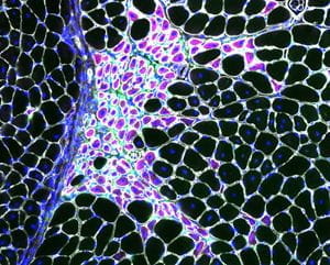
Macrophages (green) closely associated with regenerating fibers (purple) in dystrophin-deficient skeletal muscle.

Duchenne muscular dystrophy (DMD) is a devastating muscle disease caused by genetic mutations to the dystrophin gene. Without dystrophin the mechanical infrastructure of striated muscle cells is compromised causing destabilization of the muscle cell membrane and cellular necrosis. While membrane fragility contributes to the pathophysiology of DMD, it alone cannot account for many pathological features of the disease. The pathophysiology of dystrophin-deficiency is intertwined with multiple secondary defects, including perturbations in the epigenetic regulation of gene expression, as well as considerable involvement of other tissues such as the immune system and stromal cells. Patients could benefit from effective countermeasures that target events downstream of dystrophin-deficiency. Our findings have shown that ⍺Klotho is epigenetically silenced in dystrophic muscles at the onset of dystrophinopathy and that restoring ⍺Klotho expression systemically or via infiltrating bone marrow-derived cells improves regeneration and function. Ongoing investigations, supported by the Muscular Dystrophy Association, are aimed at investigating the mechanisms through which restoring ⍺Klotho reduces fibrosis in dystrophic muscles.

Other investigations in our lab include exploring the molecular mechanisms controlling the differentiation muscle mesenchymal cells (Fibro/adipogenic progenitors) and pathological fibrosis in dystrophin-deficient muscles, supported by Ralph W. and Grace M. Showalter Research Trust. The laboratory is also exploring mechanisms through which fibroblasts could promote cardiac myocyte injury, hypertrophy and remodeling in the dystrophin-deficient heart.

image showing Pathological remodeling of the dystrophin-deficient myocardium

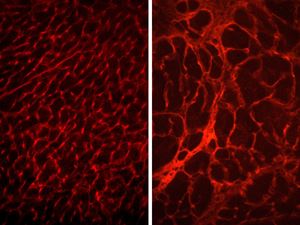
Pathological remodeling of the dystrophin-deficient myocardium (right).

Myeloid cell-mediated mechanisms of muscle regeneration

cover of the journal of immunology showing work from the welc lab

Inflammatory lesion in injured muscle featured on the cover of The Journal of Immunology (Vol. 205, Issue 6. DOI: 10.4049/jimmunol.2000247)

A major goal of the laboratory is to understand the regulatory interactions between the immune system and skeletal muscle that are important in regulating muscle regeneration. In recent years, appreciation for the contributions of myeloid cells to the regeneration of injured muscle has grown. However, our understanding of the molecular mechanisms that activate macrophages to a pro-regenerative phenotype and the specific soluble factors produced by macrophages to facilitate regeneration is incomplete. We are attempting to understand the signaling pathways that activate macrophages to an M2-biased pro-regenerative phenotype. Using a mouse line in which there is a targeted deletion of PPARδ in myeloid cells we determined that PPARδ regulates the recruitment of macrophages to injured muscle but did not reduce the expression of phenotypic markers associated with M2 macrophage activation. These findings are distinct from other notable investigations that demonstrated the importance of PPARδ expression and activation in regulating the phenotype of liver- and adipose tissue-resident macrophages and suggest that the mechanisms that regulate M2-biased macrophage activation differ in different in vivo environments. The lab is also interested in the activation of muscle macrophages to non-canonical phenotypes in muscular dystrophy and with aging, as well as potential maladaptive consequences to muscle health.

Research Team

Led by Steven Welc, PhD, research team members include Felicia Kennedy, MA, (lab manager) and Cyan Cosby, BS, (research technician).