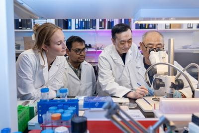
Faculty Research Labs

Gao Lab

sample of brain tissue with a curving blue line made of small dots and red neural networks moving above and below

The laboratory of Xiang Gao, PhD focuses his investigations on the mechanisms of traumatic brain injury (TBI). His research is threefold: to find an effective way to mitigate, or even prevent, cell death and dendritic/synaptic degeneration post TBI; to explore potential therapeutic interventions for repairing damaged neural circuitries; and to study the molecular mechanisms underlie immune response and neuroinflammation post-trauma.

An estimated 2.8 million incidences of TBI occur within the United States annually, and the injury contributes to a substantial number of trauma-related deaths and also permanent disabilities in survived patients that has a devastating impact on them and their families. Unfortunately, there is no effective treatment for this disease. Preventing cell death—stopping or blocking damage from the beginning of insult—is the most effective way to halt TBI. Neuronal excitotoxicity caused by robust increase of glutamate post-trauma is a primary area of interest for the Gao Lab. A glutamate signaling pathway has been proven to play a critical role in neuronal death after TBI. However, it isn’t fully understood. The Gao lab is furthering this study, and, for the first time, morphologically revealed extensive dendrite and spine degeneration in spared neurons after TBI. Live imaging using a cutting-edge thinning-skull technique and combined with 2-photon confocal microscopy further confirmed this phenomenon in vivo. The timing of dendritic and synaptic degeneration correlated with functional loss and suggested it may have a major contribution to the deficit. Moreover, dendritic and synaptic degeneration happened in a comparable later phase, about 24 hours after injury in an animal model, allow us to practically prevent it, especially in the clinic.

The Gao Lab designed a stem cell niche in the cortex using an in vivo reprogramming method. Damaged neural circuitries directly result in associated neurological disorders. Stem cells could be affected by their local environment to generate sufficient neurons to fill the cavity in the injured cortex. This method may create a potentially therapeutic application for curing TBI. Other than regenerating new neurons to compensate the loss, directly targeting and managing function-related neural circuitry and improving functional outcomes seems a promising novel intervention.

Overwhelming evidence has revealed that TBI triggers sterile neuroimmune and neuroinflammatory responses that can have both detrimental and beneficial effects. While appropriate acute and transient neuroimmune responses facilitate the repair and adaptation of injured brain tissues, prolonged and excessive neuroinflammatory responses may accelerate TBI brain damage. The mechanisms that control the intensity and duration of neuroimmune and neuroinflammatory responses in TBI largely remain elusive. The Gao lab plans to study the role of immune checkpoints in the regulation of neuroimmune and neuroinflammatory responses in the brain in vivo.

The projects in the lab are designed to study the molecular mechanisms underlying pathologies after TBI. This work will lead to identifying vital targets for clinical studies with a team of neurosurgeons at IU School of Medicine.

Active Research

  • Preventing progressive spine loss to improve function recover after repetitive mild traumatic brain injury (rmTBI)

    Youth, high school, college and professional athletes who participate in contact sports with a history of Repetitive Mild Traumatic Brain Injury (rmTBI) show increased rates of cognitive impairment, psychiatric disorders and potentially Chronic Traumatic Encephalopathy (CTE). To date, there is no FDA approved effective treatment for rmTBI, mostly due to poor understanding of the pathophysiology following rmTBI. Excitatory synapses loss has been suggested to play a major role in the etiology of rmTBI. The Gao Lab first demonstrated that single mTBI causes extensive dendritic spine loss without inducing obvious cell death and axonal damage, and it positively correlated with functional impairment. A recent study in rmTBI model in the lab also revealed dramatic spine loss after injury. The mechanisms of spine loss after mTBI are also poorly understood. The Gao Lab found that spine loss was not transient after rmTBI. Instead, it was a progressive process lasting from days to at least a week and is an acute and transient event after initial injury. Recent studies in other neurodegenerative diseases, such as Alzheimer’s disease, Parkinson’s disease and Huntington’s disease revealed the important role of inflammation in spine loss. It has been shown that microglia could engulf the spine through a process mediated by activation of a complement system, which highlighted a possible way of spine elimination in other pathological conditions, including rmTBI. Therefore, the Gao Lab proposes that engulfment by reactive microglia via activation of the complement system is one of the major mechanisms underlying the progressive spine loss after rmTBI. This inhibition may prevent spine loss and result in better functional outcomes. This study by the lab will augment their knowledge of the etiology of rmTBI, and it will highlight the complements as new targets for developing therapeutic interventions to treat the neurological disorders caused by rmTBI.

  • The function of proliferative reactive astrocyte after TBI
    Astrocytes react to brain injury with progressive changes in gene expression, morphological hypertrophy and proliferation referred as astrogliosis. The beneficial or detrimental effects of proliferative reactive astrocytes are constantly under debate, and the underlying molecular mechanisms are poorly understood. The long-term goal of the Gao Lab is to dissect the mechanisms underlying the proliferation of reactive astrocytes and its biological function in the hippocampus after TBI and in order to find the vital targets for TBI treatment. The Gao Lab is studying the role of platelet-derived growth factor receptor alpha signaling in regulating reactive astrocyte proliferation, and we believe that proliferative reactive astrocytes are neuroprotective by favoring synapse preservation and regeneration following TBI. Completion of mechanism study will provide an ideal tool to explore the function of proliferative reactive astrocyte in the hippocampus and which in turn to identify vital targets for therapeutic intervention aiming at functional improvement after TBI. This project is innovative because the lab reveals for the first time the role of the receptor in regulating reactive astrocyte proliferation post-trauma using state-of-art inducible conditional knock-out/knock-in technique and discover the neuroprotective aspects of proliferative reactive astrocytes in injured hippocampus.
  • The molecular mechanisms underlying immune responses and inflammation after TBI
    Overwhelming evidence has revealed that TBI triggers sterile neuroimmune and neuroinflammatory responses that can have both detrimental and beneficial effects. While appropriate acute and transient neuroimmune responses facilitate the repair and adaptation of injured brain tissues, prolonged and excessive neuroinflammatory responses may accelerate TBI brain damage. The mechanisms that control the intensity and duration of neuroimmune and neuroinflammatory responses in TBI largely remain elusive. The Gao Lab used TBI to study the role of immune checkpoints (ICPs) in the regulation of neuroimmune and neuroinflammatory responses in the brain in vivo. They found that de novo expression of PD-L1 (programmed death-ligand 1)—a key inhibitory ICP that exerts dual inhibitory signals to suppress the activity of PD-1-expressing T cells via the PD-1/PD-L1 axis and a novel intrinsic reverse signaling—was robustly and specifically induced in reactive astrocytes. These PD-L1-positive astrocytes, including newly generated proliferative astrocytes were highly enriched to form a dense zone around the TBI lesion. Inhibition of PD-L1 signaling suppressed the cell survive and prompted the expression of neuroinflammatory related genes, such as STAT3 and its downstream target CCL2, which resulted in increase of inflammatory Ly-6CHigh monocytes / macrophages (M/Mϕ) infiltration, brain tissue cavity size and subsequent motor and emotional dysfunction in TBI mice. The lab therefore hypothesizes that neuroprotective PD-L1 signaling is involved in astrocytic scar formation and PD-L1-positive astrocytes act as a brake to control TBI-induced neuroimmune and neuroinflammatory responses via counteracting CCL2 expression through suppressing STAT3 signaling. The research of the Gao Lab will demonstrate that PD-L1-positive astrocytes exert their dual inhibitory signals to affect the immune responses and functional outcome following TBI, thereby opening a novel avenue to study the role of ICPs in TBI pathophysiology and provides insights into development of ICP and its related signal pathway regulators to improve TBI functional outcomes.
  • The molecular mechanisms underlying great neuroprotection of Dexmedetomidine, a FDA approved sedative drug after trauma
    Systematic application of therapeutic hypothermia (TH) in the acute phase post-TBI has been shown to be neuroprotective in both experimental and human studies. Despite the discourage data from multicenter randomized clinic trails (RCT), which might be due to unsuccessfully achieve desired temperature (330C) within therapeutic time window and other limitations, it is important to continually the topic of TH and its utility in neuroprotection. The long-term goal of the Gao Lab is to find a target to improve the therapeutic effects of hypothermia by either developing strategy for on-site using or extending the therapeutic window and investigating the putative synergistic effect of combining hypothermia with other neuroprotectants. The lab's pilot study showed that in a mouse model of moderate TBI, one dose of the FDA-approved sedative Dexmedetomidine (Dex) rapidly generated long last TH (32-350C) in an ambient temperature at 24.50C. This modulation of body temperature without application of any auxiliary heating and cooling equipment suggests that Dex can be potentially used on-site. Importantly, Dex treated TBI mice with induced TH exerted greater functional improvement than conventional TH or Dex treated TBI mice with normothermia, reflecting by further reduced lesion area in cortex and better behavior test score. Dex has also been proven to activate multiple signaling pathways which can be neuroprotective. Therefore, the Gao Lab believes that Dex exerts a great neuroprotective effect on TBI via a synergistic mechanism of inducing early hypothermia and activating inherent neuroprotective signaling. This research is innovative because the lab reveals for the first time the potential role of “old” FDA approved Dex in treating TBI patients, which could be rapidly assimilated into the clinical practice.
  • The new intervention to alleviate the symptoms after TBI via managing function associated neural circuit
    TBI causes a wide range of pathological changes in the brain that lead to multiple neurological disorders. These disorders remain incurable mostly due to the lack of knowledge about the connections that wire pathological changes to the specific symptom after TBI. Recent studies and emerging new technologies have facilitated function/behavior associated neural circuitry identification. This provides an opportunity to bridge the gap between pathologies and functional outcomes after trauma. Moreover, it opens a new avenue to treat neurological disorder via directly targeting associated neural circuitry by neuronal activity management post-trauma. Risk-taking behavior is a commonly reported emotional disorder among young TBI patients and veterans with PTSD. The deficits of risk-taking behavior post-TBI have drastically negative impact on patient’s social and professional outcomes, their safety, their own and their relative’s quality of life. To date, no treatment for this devastating disorder.  The Gao Lab replicated this symptom in a controlled cortical impact (CCI) mouse model of moderate TBI and studied the alteration of its associated neural circuit following trauma. The preliminary data demonstrates the dramatic increase of risky behavior in the TBI mice, even 12 weeks post-injury. Staining with antibody against c-fos, a marker for neuronal activity, revealed that TBI significantly damaged hippocampal-hypothalamic circuit with a remarkable decrease of neuronal activity in ventral hippocampal CA1 (vCA1) region and lateral hypothalamic area (LHA). The neuronal activity in these two regions, which belong to basolateral amygdala (BLA)-ventral hippocampus CA1 (vCA1)-hypothalamus area (LHA) circuit, a direct route by which the hippocampus can rapidly influence innate anxiety behavior, has been suggested to be critical for controlling risky activity as well. Taking advantage of chemogenetic system Designer Receptors Exclusively Activated by Designer Drugs (DREADD), the lab manually increased vCA1 neuronal activity of TBI mice via overexpressing Dreadd receptor (hM3D(Gq)-mcherry, activator) in this region by adeno-associated viruses (AAV) mediated delivering. The Dreadd infected vCA1 neurons not only projected into LHA, but also enhanced the neuronal activity in LHA when they were activated by clozapine N-oxide (CNO), a specific ligand for Dreadd receptor activation. Further, the increase of neuronal activity in vCA1 and LHA may eventually lead to the mitigation of risky behavior after TBI. Taken together, the disruption of hippocampal-hypothalamic circuit is the molecular mechanism underlies risk-taking behavior after TBI and risky behavior is able to be mitigated or even prevented by neural circuit management using DREADD chemogenetic system. Successful completion of the proposed study will provide new insights to support the long term goal to better understand the mechanisms that underlie specific neurological dysfunction after TBI, a critical step to identify targets for focused interventions. Although, virus mediated chemogenetics successfully modulates the neuronal activity in vCA1 and may mitigate risk-taking behavior in mice, this procedure is too invasive for clinical use. Thus, the future studies with exploring novel noninvasive neuromodulation methods, such as using magnetic strategies for neuronal activity management are next steps aiming at achieving the same effects practically.
19839-Gao, Xiang

Xiang Gao, PhD

Assistant Research Professor of Neurological Surgery

Read Bio Xiang Gao, PhD