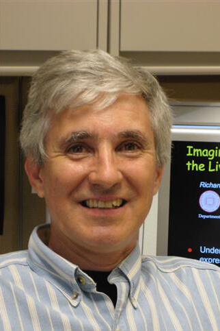
Richard N. Day, PhD, MS
Professor Emeritus of Anatomy, Cell Biology & Physiology
- Phone
- (317) 274-2166
- Address
-
MS 333
PBIO
IN
Indianapolis, IN - PubMed:
-

Bio
Research in my laboratory applies quantitative fluorescence microscopy techniques to visualize the behaviors of proteins inside living cells. Our studies use the combination of molecular biology, biochemistry, and live-cell imaging approaches to determine how specific signal transduction and gene regulatory complexes are assembled. We are using time-resolved fluorescence lifetime imaging microscopy (FLIM) to measure Förster resonance energy transfer (FRET), which enables us to determine how certain disease-causing point mutations can affect the assembly of specific protein complexes. I have over one hundred publications, with extensive experience in live-cell imaging. In addition, I have taught in the annual Cold Spring Harbor Laboratory or Marine Biological Laboratory live-cell imaging courses yearly since 1998, and I am currently the co-organizer for the Workshop on FRET microscopy, held annually at the University of Virginia since 2001.
| Year | Degree | Institution |
|---|---|---|
| 1990 | Postdoctoral Training | University of Iowa |
| 1987 | PhD | University of Rochester |
| 1980 | MS | State University of New York |
| 1978 | BA | University of Colorado |
Research
1. My early research focused on how the interactions between cell-specific transcription factors, their co-regulatory protein partners and the nuclear receptors function in concert to regulate gene expression. We used biochemical, genetic, and molecular approaches to determine how the protein complexes that are necessary for pituitary specific gene transcription are assembled. Our studies demonstrated that Pit-1, a pituitary-specific homeodomain transcription factor, orchestrates the activities of a network of transcription factors and co-regulatory proteins that function to control pituitary gene transcription. Together, these studies showed how Pit-1 interacted cooperatively with nuclear receptors and other transcription factors and coregulatory proteins to regulate prolactin (PRL) and growth hormone gene expression.
-
Day, R.N., Koike, S., Sakai, M., Muramatsu, M., Maurer, R.A. 1990. Both Pit-1 and the estrogen receptor are required for estrogen responsiveness of the rat prolactin gene. Molecular Endocrinology 4:1964-1971. PMID: 2082192.
-
Day, R.N. Day, K.H. 1994. An alternatively spliced form of Pit-1 represses prolactin gene expression. Molecular Endocrinology 8:374-381. PMID: 8152427.
-
Day, R.N., Liu, J., Sundmark, V., Kawecki, M., Berry, D., Elsholtz, H.P. 1998. Selective inhibition of PRL gene transcription by the ets domain factor, ERF. Journal Biological Chemistry 273:31909-31915. PMID: 9822660.
-
Schaufele F, Enwright JF III, Wang X, Teoh C, Srihari R, Erickson R, MacDougald OA, Day, R.N. 2001. CCAAT/enhancer binding protein alpha assembles essential cooperating factors in common subnuclear domains. Molecular Endocrinology 15:1665-1676. PMID: 11579200.
2. Importantly, we complemented these structural and functional studies with live cell imaging studies. This integrative approach allowed us to determine how the subnuclear positioning of protein complexes contributes to the selective expression of tissue-specific genes. We used fluorescence microscopy to visualize the intranuclear distribution and interactions of proteins labeled with a variety of different color fluorescent proteins. These studies in living cells showed how the assembly of cooperating factors at particular intranuclear sites is critical for the regulation of cell-specific gene expression. We used this approach to demonstrate that Pit-1 specifically recruited nuclear receptors and other transcription factors and coregulatory proteins to the nuclear sites it occupied, providing evidence that Pit-1 can direct cooperating factors to particular sites in the nucleus.
-
Day, R.N. 1998. Visualization of Pit-1 transcription factor interactions in the living cell nucleus by fluorescence resonance energy transfer microscopy. Molecular Endocrinology 12:1410-1419. (Cover Article). PMID: 9731708.
-
Enwright III, J.F., Kawecki-Crook, M.A., Voss, T.C., Schaufele, F., Day, R.N. 2003. A Pit-1 homeodomain mutant blocks the intranuclear recruitment of the CCAAT/Enhancer binding protein alpha required for prolactin gene transcription. Molecular Endocrinology 17(2):209-222. PMID: 12554749.
-
Voss, T.C., Demarco, I.A., Booker, C.F., Day, R.N. 2005. Functional Interactions with Pit-1 Reorganize Corepressor Complexes within the Nucleus. Journal of Cell Science 118: 3277-3288. PMID: 16030140.
-
Demarco, I.A., Voss, T.C, Booker, C.F., Day, R.N. 2006. Dynamic Interactions between Pit-1 and C/EBP alpha in the Pituitary Cell Nucleus. Molecular and Cellular Biology 26:8087-8098. PMID: 16908544.
3. These studies also demonstrated that the p42 CCAAT/enhancer-binding protein alpha (C/EBPα) interacted cooperatively with Pit-1 to activate PRL transcription. This was of particular interest because C/EBPa is a basic region-leucine zipper (BZip) transcription factor that functions to direct programs of cellular differentiation. C/EBPα is unusual among transcription factors in that it binds preferentially to α-satellite DNA elements located in regions of centromeric heterochromatin. Furthermore, C/EBPα requires DNA methylation to optimally bind DNA elements in gene promoters. Our published studies showed that the BZip domain of C/EBPα is necessary and sufficient for targeting to the centromeric heterochromatin in mouse cells, and critically demonstrated that the C/EBPα BZip domain directly interacts with heterochromatin protein 1 α (HP1α).
-
Demarco, I.A., Periasamy, A., Booker, C.F., Day, R.N. 2006. Monitoring Dynamic Protein Interactions with Photo-quenching FRET. Nature Methods 3:519-524. PMID: 16791209.
-
Siegel, A.P., Hays, N.M., Day, R.N. 2013. Unraveling transcription factor interactions with heterochromatin protein 1 using fluorescence lifetime imaging microscopy and fluorescence correlation spectroscopy. J Biomed Optics 18:25002. PMID: 23392382.
-
Tsekouras, K., Siegel, A.P., Day, R.N., Presse, S. 2015. Inferring Diffusion Dynamics from FCS in Heterogeneous Nuclear Environments. Biophysical journal 109 (1):7-17. PMID: 26153697.
4. Over the past decade we have pioneered the use of fluorescence lifetime imaging microscopy (FLIM) to measure Förster resonance energy transfer (FRET) between proteins labeled with FPs inside living cells. FLIM quantifies FRET by the direct measurement of the donor fluorescence lifetime alone. This eliminates problems associated with spectral bleedthrough, making FLIM among the most accurate method for measuring FRET. We have also developed the complementary approach of fluorescence correlation spectroscopy (FCS) to monitor protein dynamics. We are currently applying these live-cell imaging methods to establish how the HP1α-C/EBPα network controls epigenetic processes in living cells.
-
Sun, Y., Hays, N.M., Periasamy, A., Davidson, M.W., Day, R.N. 2012. Monitoring protein interactions in living cells with fluorescence lifetime imaging microscopy. Methods in Enzymology: Live Cell Imaging 504:371-91. PMID: 22264545.
-
Day, R.N. 2014. Measuring protein interactions using Förster resonance energy transfer and fluorescence lifetime imaging microscopy. Methods 66(2):200-207. PMID: 23806643.
-
Siegel, A.P., Baird, M.E., Davidson, M.W., Day, R.N. 2013. Strengths and Weaknesses of Recently Engineered Red Fluorescent Proteins Evaluated in Live Cells Using Fluorescence Correlation Spectroscopy. Int. J. Mol. Sci. 14(10):20340-20358. PMID: 24129172.
-
Day, R.N. 2015. Measuring Förster Resonance Energy Transfer Using Fluorescence Lifetime Imaging Microscopy. Microscopy Today 23(3):44-50.
The research in the Day laboratory has been funded by the National Institutes of Health, and we gratefully acknowledge the support of the Indiana University School of Medicine, and the past support of the NSF, the NSF Center for Biological Timing, and the American Cancer Society.