3D Bioprinting at IU School of Medicine

A transplant is often the only option for a patient’s recovery when their organs fail. From enlarged hearts to livers ridden with disease, many people find themselves waiting months or even years for a transplant that will save their life. However, despite the great need for donation, there is still a shortage in organ donors, leaving more than twenty people a day in the United States who die waiting for organ donation. To help meet the growing need for donation, experts at IU School of Medicine are working to advance research in xenotransplantation using 3D bioprinting.

Growing Cells and Growing Hope
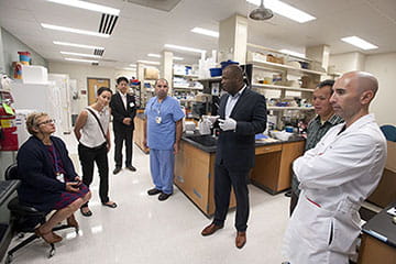
Experts at IU School of Medicine are one of two institutions to use Regenova 3D bioprinting technology. Adapted by Cyfuse Biomedical, Regenova is operated with a Windows machine using custom software which permits customized tissue construct design and automated cell placement. This system relies on a scaffold-free method to generate tissue constructs using the Kenzan Method. In this method, extremely dense sphere cell materials are skewered onto an array of needles. Over time, the cells begin to fuse and form a solid structure, where they are then placed in a bioreactor.

This advanced technology will enable researchers at IU School of Medicine and beyond to set the stage for future advancements toward the growing demand for organ donation.

110000 people in the US waiting for a life-saving organ transplant
9 million awarded to further 3D bioprinting research at IU School of Medicine
20 people die daily waiting for a transplant

Next Generation Printing

Cell placement takes place in a closed cabinet protected by a high efficiency particulate air filter, which manages cell spheroid contamination and aerosol exhaust. Plates containing spheroids are loaded into the two right sides and then are loaded into the left side once all cells have been removed.

Kenzan Method

Due to cell thickness and its lack of elasticity, previous attempts of generating tissue from cultured cells has proven challenging. The Kenzan Method focuses on natural features of cell self-aggregation to assemble spheres of cells into any desired three-dimensional macroscopic tissue without collagen or hydrogel materials, allowing for easier circulation during the culture period. As the cells are cultured and placed in fine needle arrays, it allows them to merge with adjacent cells. With the needles appropriately aligned, cells can be positioned in any desired three-dimensional layout. Finally, culturing connected cells in a bioreactor yields a 3D tissue with a desired function and quality.

Quick Facts

  • What is bioprinting?
    3DĀ bioprintingĀ is a form of automated tissue engineering which produces cell-based structures useful for a variety of in vitro and in vivo applications.
  • Is it available for clinical trials?
    Not yet, but with this important research at IU School of Medicine, experts are paving the way toward clinical trials.

Bioprinting Experts

Burcin Ekser, MD, PhD

Associate Professor of Surgery

Lester J. Smith, PhD

Assistant Professor of Radiology & Imaging Sciences